


Digital X-ray machines are diagnostic imaging systems that use X-rays to record internal body structures and convert the acquired data into digital images. Compared with conventional film-based X-ray systems, digital technology allows images to be processed, stored, retrieved, and shared through healthcare information systems.
Among current solutions, DR (Digital Radiography) systems are of particular interest to many healthcare facilities because they acquire digital images using dedicated detectors and can support a more convenient imaging and image-management workflow.
However, investing in a digital X-ray system involves more than choosing equipment with high resolution or fast image-display times. Hospitals and clinics should also evaluate detector technology, image quality, radiation-exposure management, system configuration, X-ray room requirements, and integration capability with PACS/RIS.
This article explains the principles of DR technology, the differences between DR and CR, the main technical components, and the criteria healthcare facilities should consider when investing in a digital X-ray system.
What Is a Digital X-Ray Machine?

A digital X-ray system is an imaging system that uses X-rays, with the acquired signals converted into digital data for processing and display on a computer.
In principle, X-rays are generated by the X-ray tube and pass through the anatomical area being examined. Different tissues and structures attenuate the X-ray beam to different degrees. The image receptor detects the transmitted X-ray signal, allowing the system to generate an image for professional assessment.
Two common technologies in digital radiography are:
- CR (Computed Radiography): uses an imaging plate capable of storing exposure information and requires a separate image-reading system.
- DR (Digital Radiography): uses a digital image detector, commonly a flat-panel detector, to transfer image data to the processing system without requiring the cassette-scanning step used in CR.
Both technologies generate digital images, but they differ in image-acquisition methods, processing, and workflow organization.
Is a DR X-Ray System a Digital X-Ray System?
Yes. DR is one technology within the broader category of digital X-ray systems.
In procurement, the term “digital X-ray machine” is often used to refer to DR systems. Technically, however, CR is also a form of digital radiography.
Therefore, when reviewing quotations or technical documentation, facilities should confirm whether the system uses a DR detector or a CR system rather than relying only on the commercial product name.
1. How Do DR and CR Technologies Differ?
The key difference between CR and DR lies in how X-ray signals are recorded and converted into digital images.
CR Technology (Computed Radiography)
A CR system uses a cassette containing a specialized imaging plate, commonly based on photostimulable phosphor material.
After exposure, the cassette is inserted into a CR reader. The system scans the stored signal on the imaging plate, converts it into digital data, and sends the image to processing software.
The basic workflow includes:
- X-rays pass through the anatomical area being examined.
- The CR imaging plate stores the exposure information.
- The cassette is inserted into a dedicated reader.
- The data are converted into a digital image.
- The image is processed, displayed, and stored.
One advantage of CR is that it may allow continued use of certain existing X-ray systems if the configuration is compatible.
However, handling the cassette and transferring it to the reader adds an additional step to the image-acquisition workflow.
DR Technology (Digital Radiography)
DR systems commonly use a flat-panel detector (FPD) to receive the X-ray signal.
The signal is converted into electronic data and transmitted to the image-processing system.
Depending on the design, the detector may be integrated into the examination table, installed in a wall stand, or used as a portable detector panel.
The basic workflow includes:
- The system generates X-rays according to the established exposure conditions.
- The DR detector receives the X-ray signal.
- The detector converts the signal into electronic data.
- The software processes and displays the image.
- The image is reviewed and sent to the storage system.
DR does not require the cassette-reading step used in CR. This can simplify image-acquisition workflow, particularly in facilities with high examination volumes.
Comparison of DR and CR X-Ray Systems
| Criterion | CR Technology | DR Technology |
|---|---|---|
| Acquisition method | Imaging plate inside a cassette | Digital image detector, commonly a flat-panel detector |
| Image-reading step | Requires a CR reader | Does not require a CR cassette reader |
| Processing workflow | Includes cassette-transfer steps | Data are transmitted from the detector |
| Image-display time | Depends on cassette-reading workflow | Generally more convenient because intermediate steps are reduced |
| Ability to upgrade an existing system | May allow continued use of a compatible X-ray generator | May be implemented as a retrofit or new installation depending on configuration |
| Maintenance | Reader, cassette, and related components | Detector, connectivity, software, and related components |
| Investment cost | Depends on the solution and existing infrastructure | Depends on the number of detectors and system configuration |
| Image quality | Depends on technology and conditions of use | Depends on the detector, X-ray generation system, and image processing |
It should not be assumed that every DR system produces better images than every CR system under all conditions. Image quality should be evaluated using the specific system configuration and the facility's quality-control program.
In addition, post-processing capability does not mean that the system can correct every exposure error or replace the need to acquire an appropriate signal from the beginning.
2. Components of a DR Digital X-Ray System
A complete DR system typically consists of multiple components operating together.
| Component | Primary Role |
|---|---|
| X-ray tube | Generates the X-ray beam |
| High-voltage generator | Provides electrical power and controls X-ray generation |
| Collimator | Limits the radiation field |
| DR detector | Acquires the X-ray signal and creates digital data |
| Examination table and wall stand | Support patient positioning and image-acquisition positioning |
| Mechanical system | Adjusts the position of system components according to the design |
| Image-acquisition workstation | Manages image acquisition, processing, and review |
| Software and data connectivity | Manage information, image transfer, and system integration |
Not all DR X-ray systems have the same configuration. Fixed systems, mobile systems, and retrofit solutions for existing X-ray equipment may differ in the number and arrangement of components.
X-Ray Tube and High-Voltage Generator
The X-ray tube generates the radiation used for image acquisition.
This operation is controlled by the high-voltage generator and other system-control components.
When evaluating the equipment, facilities should review:
- Generator power and operating range.
- X-ray tube characteristics.
- Heat-load management capability.
- System stability.
- Protection and alarm functions.
- Availability of spare parts and maintenance services.
The system should not be selected solely on the basis of the highest nominal generator output.
Facilities should determine which power level and configuration are appropriate for their imaging needs, utilization frequency, and actual operating conditions.
Collimator and Positioning System
The collimator limits the X-ray field to the area that needs to be imaged.
The positioning system may include:
- Examination table.
- Detector stand.
- X-ray tube column or ceiling-mounted support.
- Position-adjustment mechanisms.
- Mechanical locking and protective components.
The ability to adjust and coordinate the X-ray tube, radiation field, and detector affects operational convenience.
For facilities serving different patient groups, accessibility to the examination table, height adjustment, and equipment movement space should also be considered.
3. How Does a DR Detector Work?

The DR detector is one of the most important components of a digital X-ray system.
It receives the X-ray signal and converts it into electronic data so that software can generate an image.
In flat-panel detector systems, two conversion methods are commonly discussed: indirect conversion and direct conversion.
Indirect-Conversion Detector
An indirect-conversion detector uses a scintillator layer to convert X-rays into light, after which the sensor system converts the light into an electrical signal.
Common materials include:
- Cesium iodide (CsI).
- Gadolinium oxysulfide (GOS or Gd₂O₂S).
Depending on the design, the detector may use an appropriate sensor matrix and signal-readout circuitry.
The material characteristics, scintillator structure, and sensor system all influence detector performance.
Direct-Conversion Detector
A direct-conversion detector uses a material capable of converting X-ray energy into electrical charge without the intermediate light-conversion stage used in indirect detectors.
One example of a material used in direct-conversion technology is amorphous selenium (a-Se).
It is important to note that the term Digital Radiography (DR) does not mean that every DR detector uses direct conversion.
DR systems may use either direct- or indirect-conversion detectors.
Specifications to Review When Selecting a Detector
| Specification | Relevance to Evaluation |
|---|---|
| Acquisition area size | Affects the anatomical coverage that can be recorded in a single exposure |
| Pixel pitch | Related to spatial sampling |
| Image matrix | Number of pixels in the acquired data |
| MTF | Characterizes contrast-transfer performance across spatial frequencies |
| DQE | Characterizes the detector's efficiency in using X-ray quantum signal information |
| Signal-response range | Related to the ability to record signals across an exposure range |
| Mechanical durability | Important for portable detectors that are frequently handled or removed |
| Connectivity | Wired or wireless connectivity and compatibility with the workstation |
| Maintenance conditions | Calibration, repair, battery replacement, and detector replacement capability |
Is DQE an Important Image-Quality Metric?
DQE (Detective Quantum Efficiency) describes how effectively a detector preserves signal information relative to input quantum noise under specified measurement conditions.
IEC 62220-1-1:2015 specifies a method for determining DQE for detectors used in radiographic imaging.
However, a single DQE value should not be compared between two products without understanding the measurement conditions, spatial frequency, and system characteristics.
Similarly, a detector with a higher megapixel count does not automatically provide better diagnostic image quality in every situation.
4. Common Digital X-Ray System Configurations
DR systems can be designed in different configurations to support different operating environments.
Fixed DR X-Ray System
This type of system is installed in a dedicated X-ray room.
The configuration may include:
- Examination table.
- Wall stand.
- X-ray generator and tube.
- One or more detectors.
- Image-processing workstation.
- Mechanical and control components.
This configuration may be suitable for facilities that need routine imaging operations in a dedicated room.
When selecting a system, facilities should consider space, positioning capability, level of system integration, and the number of detectors.
Mobile Digital X-Ray System
A mobile DR X-ray system is designed to be moved to appropriate locations within its stated intended use.
The equipment may be considered for intensive-care units, emergency departments, or other areas where bedside imaging is required according to professional indications.
Important criteria include:
- Size and weight.
- Mobility.
- Battery or power supply.
- Operational stability.
- Detector protection.
- Image and data-management capability.
- Radiation-safety conditions in the area of use.
Mobile systems are not exempt from radiation-protection requirements simply because they are not permanently installed in a dedicated X-ray room.
Retrofitting an Existing X-Ray System to DR
Some facilities may consider installing a DR detector and processing software to upgrade an existing X-ray system.
This approach is commonly referred to as a DR retrofit.
However, upgrade feasibility depends on:
- Condition of the existing generator and X-ray tube.
- Ability to synchronize the X-ray exposure signal with the detector.
- Control interface.
- Mechanical system.
- Electrical and radiation-safety condition.
- Software compatibility.
- Post-upgrade testing requirements.
Replacing the detector does not mean that the entire existing system has become a new X-ray system with equivalent performance and safety characteristics.
Facilities should separately evaluate whether the existing components can continue to be used and clarify maintenance responsibilities among suppliers.
5. Image Quality and Radiation-Exposure Management
One advantage of digital radiography is the ability to process and adjust image presentation after acquisition.
However, this also creates a need for tighter technical control.
Does a Clear Image Mean the Exposure Level Is Appropriate?
No.
DR systems may generate images with relatively stable displayed brightness and contrast across a wide exposure range.
Therefore, simply looking at whether an image appears bright or dark on the monitor is not sufficient to assess exposure level.
Without an appropriate quality-control program, a system may experience dose creep — a gradual increase in exposure over time without a proportional benefit in image information.
This is why facilities should pay attention to exposure indicators, image-quality control, and analysis of repeat examinations.
EI and DI in Digital Radiography
Some DR systems provide:
- Exposure Index (EI): an index related to the exposure level at the image receptor according to a defined method.
- Target Exposure Index (EIₜ): a target value established for an appropriate imaging procedure.
- Deviation Index (DI): an index indicating the difference between the obtained EI and the target value.
These indicators can help personnel monitor whether the image-acquisition process is operating within an appropriate range.
It is important to note that EI is not a direct measurement of the radiation dose received by the patient.
Dose assessment also depends on multiple other factors, including beam characteristics, anatomical region, imaging geometry, and appropriate dosimetric indicators.
According to the ACR–AAPM–SIIM–SPR guidance on digital radiography, facilities should establish a program to evaluate exposure indicators and image quality appropriately.
Role of Automatic Exposure Control
Some X-ray systems include Automatic Exposure Control (AEC).
AEC may support termination of the X-ray exposure based on established signals and control conditions.
However, AEC should be configured, tested, and used appropriately with the image-acquisition system.
The presence of AEC should not be assumed to automatically ensure the lowest possible radiation dose or optimal image quality in every case.
6. Radiation-Safety Requirements for DR X-Ray Installation
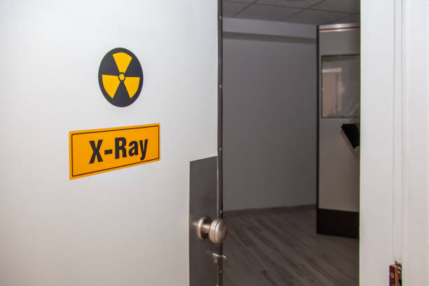
Although DR uses digital technology, the equipment still produces ionizing radiation.
Installation and operation must therefore comply with the radiation-safety requirements applicable to healthcare facilities using medical X-ray equipment.
X-Ray Room and Shielding Assessment
Before installing a fixed system, facilities should assess:
- Location of the X-ray room.
- Room size and equipment layout.
- Walls, doors, and shielding structures.
- Control area.
- Possible directions of the X-ray beam.
- Adjacent areas occupied by staff or other people.
- Ability to observe and communicate with the patient.
- Other relevant safety conditions.
According to IAEA Safety Guide SSG-46, X-ray room design should consider the type of equipment, workload, beam direction, occupancy of adjacent areas, and other radiation-protection factors.
A single fixed thickness of lead or shielding material should not be applied to every X-ray room. Shielding requirements should be calculated and evaluated for each individual installation.
Testing and Acceptance Before Use
Facilities should establish a plan for:
- Equipment-quality testing after installation.
- Radiation-safety assessment.
- Testing of the X-ray generation system.
- Radiation-field and alignment checks.
- Detector-quality assessment.
- Testing of alarm and protective functions.
- Establishing reference values for periodic quality control.
These testing activities should be performed by personnel or organizations with appropriate competence.
According to the IAEA, a quality-assurance program should include acceptance testing, commissioning evaluation, and periodic quality control.
Relevant IEC Standard
IEC 60601-2-54:2022 specifies particular requirements for the basic safety and essential performance of X-ray equipment used for radiography and radioscopy within the scope of the standard.
This standard does not generally apply to all imaging equipment categories such as CT, mammography, or dental X-ray systems.
When selecting a DR system, facilities may ask suppliers to provide documentation demonstrating conformity with relevant technical standards.
Note: The IEC standards and IAEA guidance mentioned above are international technical references. Installation, licensing, verification, radiation surveys, and operation of equipment in Vietnam should comply with the current requirements applicable to the healthcare facility.
7. Connecting Digital X-Ray Systems with PACS and RIS
One operational advantage of DR systems is their ability to integrate with medical image-data management infrastructure.
However, the ability to generate digital images does not mean that a system can automatically connect to every PACS.
What Are the Roles of PACS and RIS?
PACS (Picture Archiving and Communication System) is used to store and communicate medical images.
RIS (Radiology Information System) supports information management and workflow within the radiology department.
DICOM (Digital Imaging and Communications in Medicine) is a standard used to represent and exchange medical imaging data together with related information.
A data workflow may be organized as follows:
- Patient and examination-order information is managed within an appropriate system.
- The X-ray system receives a worklist through supported connectivity functions.
- Personnel acquire and review the images.
- Images are sent to PACS.
- Physicians retrieve images from the system for professional review.
- Results and related data are managed according to the facility's workflow.
DICOM Functions to Evaluate
| Function | Role |
|---|---|
| DICOM Storage | Transfers and stores image data in a compatible system |
| Modality Worklist (MWL) | Receives worklist information |
| Modality Performed Procedure Step (MPPS) | Communicates procedure status where supported |
| DICOM Print | Supports printing through compatible systems |
| Radiation Dose Structured Report (RDSR) | Communicates radiation-exposure event data where supported |
Not every DR system includes all of these functions by default.
Facilities should request the DICOM Conformance Statement, which describes the DICOM functions supported by the device.
Issues to Check When Integrating with PACS
- Which DICOM functions does the system support?
- Are additional software licenses required?
- Can the device receive a Worklist from RIS?
- How long are images stored locally?
- Can data be resent after a network interruption?
- Are patient-information fields managed consistently?
- Are security, user-access control, and activity logs supported?
- Which supplier is responsible for resolving integration problems?
Having a LAN or Wi-Fi connection does not mean that the system can fully integrate with the facility's existing PACS/RIS.
Hospitals should verify compatibility during site assessment and acceptance rather than waiting until after installation.
8. Quality Control and Maintenance of DR X-Ray Systems

Digital X-ray systems should be monitored throughout their service life to maintain image quality and operational safety.
According to IAEA guidance on quality assurance in radiography, the quality-control program should monitor the stability of the X-ray generation system, image detector, and related functions.
Testing the X-Ray Generation System
Items may include:
- Generator stability.
- Exposure parameters according to appropriate test methods.
- Accuracy of the control system.
- X-ray beam alignment.
- Field-limitation function.
- Protective mechanisms.
Detector and Image-Quality Testing
Items that may need to be assessed include:
- Image uniformity.
- Defective pixels.
- Noise and image lag.
- Calibration capability.
- Signal stability.
- Image-quality indicators using an appropriate method.
If wireless detectors are used, the facility should also monitor battery condition, connectivity, and storage conditions.
Reject and Repeat Analysis
Analyzing rejected or repeated images may help facilities identify factors that reduce operational efficiency.
Examples include:
- Incorrect positioning or acquisition area.
- Motion-related image artifacts.
- Connectivity or data errors.
- Inadequate signal quality.
- Equipment- or workflow-related issues.
However, this analysis should be performed within an appropriate quality-management program rather than relying only on the repeat rate.
Technical Service Requirements
When selecting a supplier, facilities should confirm:
- Availability of inspection and maintenance support in Vietnam.
- Response time when faults occur.
- Conditions for X-ray tube replacement.
- Ability to repair or replace the detector.
- Software-update arrangements.
- Post-repair quality-testing services.
- Warranty conditions for individual components.
In particular, the detector and X-ray tube may have a significant impact on long-term maintenance costs.
Therefore, total cost of ownership should be evaluated rather than focusing only on the initial purchase price.
9. Ten Criteria for Selecting a Digital X-Ray System for a Healthcare Facility
When comparing DR systems, hospitals and clinics may use the following evaluation framework:
| Criterion | What Should Be Evaluated? |
|---|---|
| 1. Intended use | Routine imaging, fixed-room imaging, mobile use, or system upgrade |
| 2. Detector technology | Detector type, size, signal quality, and durability |
| 3. X-ray generation system | X-ray tube, generator, operating capability, and maintenance |
| 4. Mechanical configuration | Examination table, wall stand, positioning, and accessibility |
| 5. Image quality | Technical indicators, image processing, and quality-control capability |
| 6. Exposure management | EI/DI indicators, AEC where available, dose information, and control functions |
| 7. Radiation safety | X-ray room conditions, shielding, acceptance, and testing |
| 8. Data integration | DICOM, PACS, RIS, storage, and information management |
| 9. Technical service | Installation, training, maintenance, X-ray tube, and detector support |
| 10. Lifecycle cost | Equipment, infrastructure, software, spare parts, and maintenance |
Common Investment Mistakes to Avoid
Comparing only detector megapixel count: Failing to evaluate DQE, MTF, stability, and actual image quality.
Assuming DR always reduces radiation dose: Overlooking exposure-management and image-quality control programs.
Failing to check PACS integration: Additional software costs or difficulties connecting with hospital systems may arise.
Focusing only on equipment price: Ignoring detector repair, X-ray tube replacement, and maintenance costs.
Overlooking X-ray room requirements: Failing to assess shielding, electrical power, space, and installation conditions before signing the contract.
Retrofitting an old system without a complete assessment: Replacing the detector without evaluating the safety and performance condition of the existing components.
10. Questions to Ask DR X-Ray Suppliers
Before purchasing, hospitals and clinics should clarify:
- Which DR technology does the system use, and what are the detector's technical characteristics?
- Is the equipment supplied as a fixed system, mobile system, or retrofit solution?
- What intended-use range is supported by the X-ray tube and high-voltage generator?
- Which specifications and test methods are used to evaluate detector quality?
- Does the system support exposure indicators and image-quality control functions?
- What requirements apply to the X-ray room, electrical power, and radiation safety?
- Which DICOM functions are supported, and can the system connect to the existing PACS/RIS?
- Does the system support temporary local data storage if the network connection is interrupted?
- Does the supplier provide installation, acceptance, training, and maintenance support?
- How are long-term costs for detector replacement, X-ray tube replacement, software, and spare parts determined?
Facilities should ask suppliers to demonstrate image acquisition, processing, and transfer to a compatible system, while also clarifying how image-quality and technical-safety acceptance will be performed before the system is placed into operation.
Frequently Asked Questions About Digital X-Ray Machines

Are Digital X-Ray Machines and DR X-Ray Machines the Same?
DR is one technology within the digital X-ray category. CR systems also generate digital images but use cassettes and dedicated image readers.
How Does a DR X-Ray System Differ from CR?
CR requires the cassette to be inserted into a reader to digitize the data, while DR uses a digital detector to transfer the signal directly to the processing system without requiring a CR cassette reader.
Does DR Provide Better Image Quality Than CR?
DR may provide workflow and image-processing advantages. However, image quality depends on the detector, X-ray generation system, exposure conditions, and quality-control program.
Do Digital X-Ray Systems Use Less Radiation Than Conventional Systems?
This cannot be concluded in every case. Detector technology and workflow optimization may help use radiation more efficiently, but exposure levels still need to be monitored and evaluated using appropriate methods.
Can a DR X-Ray System Connect to PACS?
Many DR systems support PACS connectivity through DICOM. However, facilities should check the supported functions, software configuration, and integration requirements of the specific model.
Can an Existing X-Ray Machine Be Upgraded to DR?
Some systems may be upgraded using a compatible DR detector and software. However, the condition of the X-ray generator, control system, mechanical components, safety status, and post-upgrade acceptance requirements should be evaluated.
Do Digital X-Ray Systems Require Periodic Maintenance?
Yes. The system should be managed through an appropriate inspection and maintenance program covering the generator, X-ray tube, detector, safety functions, and image quality.
Explore Digital X-Ray Technology and Diagnostic Imaging Solutions at Pharmedi Vietnam 2027
For hospitals and clinics upgrading their diagnostic imaging systems, selecting a digital X-ray system involves more than simply moving from CR to DR. Facilities should evaluate detector quality, radiation-exposure management, X-ray room requirements, and data connectivity with PACS/RIS together.
Pharmedi Vietnam 2027, taking place from September 28–30, 2027 at the Saigon Exhibition and Convention Center (SECC), Ho Chi Minh City, provides an opportunity for equipment suppliers, technical professionals, and healthcare organizations to meet and discuss technologies and solutions that support healthcare delivery.
For facilities planning to invest in a DR X-ray system, the exhibition provides an opportunity to explore developments in diagnostic imaging equipment, evaluate supplier capabilities, and prepare technical requirements before selecting a solution.
When exploring digital X-ray technology, facilities may focus on:
- Is the DR detector technology appropriate for the required examinations and expected image quality?
- Do the X-ray generation system, examination table, and wall stand meet actual operating requirements?
- Which exposure-management and quality-control functions does the equipment support?
- Can the system integrate with DICOM, PACS, and RIS within the existing information infrastructure?
- Does the supplier support X-ray room assessment, installation, acceptance, and maintenance?
- What are the long-term costs of investment, operation, and replacement of major components?
Preparing these criteria in advance helps hospitals and clinics evaluate diagnostic imaging solutions more effectively and select a configuration aligned with their infrastructure and development plans.
For digital X-ray systems, an effective investment should support image quality, radiation safety, data connectivity, and stable performance throughout the equipment lifecycle.
Dates: September 28–30, 2027
Venue: Saigon Exhibition and Convention Center (SECC), Ho Chi Minh City
Exhibition Information: https://pharmed.vn/
Product Categories: https://pharmed.vn/exhibitors/products
Visitor-registration information, participating-company lists, and activity programs will be updated according to official announcements from the Pharmedi Vietnam 2027 Organizing Committee.
This article is provided for informational purposes regarding the technological principles, components, and selection criteria for digital X-ray systems. It does not provide guidance on imaging indications, X-ray exposure settings, or interpretation of images for diagnosis. Use, testing, acceptance, and operation of the system should be performed by appropriately qualified personnel in accordance with manufacturer instructions and current radiation-safety requirements.