


A CT scanner is a diagnostic imaging system that uses X-rays and data-processing technology to reconstruct cross-sectional images of internal body structures. Unlike conventional X-ray imaging, CT systems acquire data from multiple projection angles, allowing images to be generated as individual slices and reconstructed in multiple planes.
Computed Tomography (CT) is now widely used in hospitals and diagnostic imaging centers. CT technology has evolved from systems that acquired individual slices to multidetector CT, dual-source CT, and systems using photon-counting detectors.
However, investment in a CT scanner should not be based only on the advertised number of slices or scanning speed. An appropriate system should simultaneously meet requirements for image quality, radiation-exposure management, throughput, electrical and cooling infrastructure, data connectivity, and long-term maintenance capability.
This article focuses on the operating principles, system components, image-acquisition technologies, and technical criteria that should be evaluated when selecting a CT scanner for a hospital.
What Is a CT Scanner?

A CT scanner is a diagnostic imaging system that uses X-ray radiation to acquire X-ray attenuation data from multiple angles around the anatomical area being examined.
The computer processes this data using reconstruction algorithms to generate cross-sectional images.
Using CT data, software may support:
- Image reconstruction in different planes.
- Visualization of anatomical structures on individual slices.
- Multiplanar reconstruction (MPR).
- Three-dimensional (3D) reconstruction.
- Measurement and image processing using supported functions.
- Storage, retrieval, and transfer of data to PACS.
Not all CT systems provide the same acquisition or image-processing capabilities. Advanced reconstruction functions may depend on the hardware configuration, software, and licensing.
How Does CT Differ from Digital X-Ray?
Both CT and digital radiography (DR) use X-rays, but they differ in acquisition methods and image-data structure.
| Criterion | DR X-Ray System | CT Scanner |
|---|---|---|
| Acquisition principle | Records projection images of structures on an image receptor | Acquires data from multiple angles around the region of interest |
| Image data | Primarily 2D projection images | Cross-sectional and volumetric data |
| Main configuration | X-ray tube, generator, DR detector, examination table or wall stand | Gantry, X-ray tube, detector array, CT table, reconstruction system |
| Image processing | Processing and display of digital radiographic images | Slice, multiplanar, and 3D reconstruction |
| Infrastructure requirements | Depend on the X-ray room configuration | Usually require detailed assessment of electrical power, cooling, structural load, and space |
| Radiation management | Image-quality and exposure management | Image-quality control, CTDIvol, DLP, and related indicators |
CT should not be considered simply a higher-resolution version of DR X-ray. The two systems are designed for different imaging methods and professional requirements.
1. Operating Principle of a CT Scanner
The basic principle of CT is based on acquiring X-ray attenuation data as the beam passes through different structures.
During acquisition, the X-ray tube and detector system are arranged to measure signals from multiple angles.
The acquired data are then processed to reconstruct cross-sectional images.
This process can be divided into four technical stages.
X-Ray Generation and Signal Acquisition
The X-ray tube generates a beam that passes through the anatomical area being examined.
Different tissues and materials attenuate X-rays according to their characteristics. The detector array records the transmitted signal and converts it into electronic data.
Multi-Angle Data Acquisition
In modern CT systems, the X-ray tube and detector assembly rotate inside the gantry to acquire data from multiple directions.
Depending on the acquisition method, the CT table may remain stationary or move according to a predefined program.
Image Processing and Reconstruction
Data from the acquisition system are transferred to the processing system.
Reconstruction algorithms use this data to generate CT images.
Reconstruction technologies may include:
- Filtered Back Projection (FBP): image reconstruction using filtered back projection.
- Iterative Reconstruction: iterative reconstruction designed to improve noise characteristics and image quality according to the algorithm.
- Deep Learning Reconstruction: reconstruction using deep-learning models, depending on the design and scope of the software.
It should not be assumed that all reconstruction algorithms provide equivalent results or can replace one another under every condition.
Image Display and Data Storage
After reconstruction, images are displayed on a workstation.
Depending on the system, data may undergo additional processing to create multiplanar images, volumetric images, or other supported display formats.
Information is commonly stored and exchanged through solutions compatible with DICOM and PACS.
2. Main Components of a CT System
A complete CT system consists of multiple mechanical, electronic, and software components working together.
| Component | Primary Function |
|---|---|
| Gantry | Houses the rotating assembly and image-acquisition components |
| X-ray tube | Generates the X-ray beam used for data acquisition |
| High-voltage generator | Supplies electrical energy to the X-ray tube |
| Detector array | Records X-ray signals |
| Data Acquisition System (DAS) | Converts and transfers signal data |
| CT table | Supports and moves the patient according to the system design |
| Image-reconstruction system | Processes data and generates cross-sectional images |
| Control workstation | Manages acquisition, image display, and result storage |
| Cooling system | Controls the temperature of relevant system components |
| Electrical and auxiliary systems | Provide power, protection, and operational support |
According to Siemens Healthineers training materials, CT hardware may also include power-distribution cabinets, motion-control systems, patient communication devices, and cooling-support components.
Gantry and Rotating Assembly
The gantry is the ring-shaped structure that houses components involved in CT data acquisition.
Depending on the design, the gantry may include:
- X-ray tube.
- Detector.
- High-voltage generator.
- Motion-control system.
- Collimator and beam filter.
- Cooling system.
- Data- and power-transmission components.
Some systems use slip-ring technology to support power and signal transmission between rotating and stationary components, enabling continuous rotation.
When evaluating the gantry, facilities should consider aperture diameter, accessibility, rotation speed, mechanical design, and operating limitations.
X-Ray Tube and High-Voltage Generator
The X-ray tube is a component exposed to substantial heat loads during operation.
Its operating capability depends on:
- Design and power capacity.
- Focal-spot characteristics.
- Heat-storage and heat-dissipation capability.
- Operating workload.
- Control system.
- Cooling conditions.
When purchasing a CT scanner, hospitals should not compare only stated tube power or heat-load capacity.
They should also evaluate service life under expected operating conditions, tube-replacement costs, warranty terms, and technical-support capability.
Detector Array and Data Acquisition System
The detector records the X-ray signal transmitted through the region being examined.
In multidetector CT systems, detector elements are arranged in multiple rows along the longitudinal axis of the scanner.
Detector configuration affects:
- Coverage along the body axis.
- Slice-generation capability.
- Spatial resolution.
- Acquisition speed.
- Certain specialized imaging functions.
The Data Acquisition System (DAS) receives signals from the detectors and converts them into data for reconstruction.
CT system quality should not be assessed only by the advertised number of detectors or slices.
CT Table
The CT table supports patient positioning and movement according to operational requirements.
Specifications to review include:
- Weight capacity.
- Travel range.
- Positioning accuracy.
- Height and accessibility.
- Supporting accessories.
- Ease of cleaning and maintenance.
For facilities serving different patient groups, accessibility and weight limits should be confirmed before investment.
3. Common CT Image-Acquisition Technologies
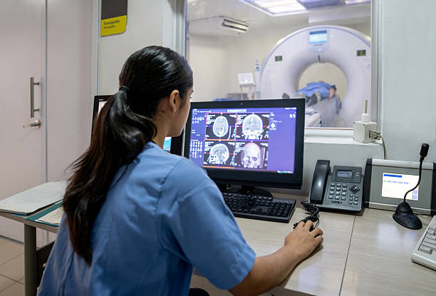
CT technology can be classified according to the data-acquisition method, detector architecture, and additional processing capabilities.
Axial CT
In sequential acquisition, the system records data at individual positions or groups of slices, after which the table may move to the next position.
This is one of the fundamental CT acquisition methods and may still be supported by modern systems.
Helical/Spiral CT
In helical CT, the X-ray tube and detector assembly rotate continuously while the table moves along the scanner axis.
Data are acquired along a helical path relative to the body.
This technology supports continuous volumetric acquisition, although image quality and radiation exposure also depend on equipment design, reconstruction method, and other professional factors.
Multidetector CT (MDCT)
Multidetector CT (MDCT) uses multiple detector rows to acquire data from several positions along the body axis during the same rotation.
Systems on the market may be described as 16-slice CT, 64-slice CT, or 128-slice CT.
However, the nominal slice count is not always equal to the number of physical detector rows.
Some systems use particular sampling or reconstruction methods to generate an output slice count that differs from the actual number of acquisition rows.
Therefore, when comparing systems, facilities should check:
- Number of physical detector rows.
- Number of acquisition channels.
- Z-axis coverage per rotation.
- Supported slice thickness.
- Rotation time.
- Image-reconstruction capability.
- Confirmed clinical applications.
Dual-Energy CT
Dual-Energy CT uses information from different X-ray energy spectra to support material-characterization analysis.
The technology may be implemented using several methods, such as:
- Two X-ray sources.
- Switching X-ray tube voltage.
- Detectors capable of separating energy information.
- Other vendor-specific spectral acquisition configurations.
Dual-Energy CT does not necessarily mean that the system has two X-ray tubes.
When evaluating this technology, facilities should confirm the acquisition method, spectral-analysis functions, and model-specific application limitations.
Photon-Counting CT
Photon-Counting CT uses detectors designed to record individual X-ray photon events and energy information.
Unlike many conventional CT detectors that use energy-integrating mechanisms, photon-counting technology may support different signal-processing and imaging approaches.
However, this technology group has specific technical and cost requirements that should be evaluated separately.
It should not be assumed that photon-counting systems are always more suitable or provide a better return on investment than every conventional CT configuration.
4. Comparing CT Configurations: Should Selection Be Based on Slice Count or Actual Needs?
When selecting a CT scanner, slice count is often one of the first specifications considered.
However, it represents only one part of the system's acquisition capability.
| Common Configuration Group | What Should Be Evaluated? |
|---|---|
| 16-slice CT | Ability to support the intended technical service portfolio, throughput, and acquisition range |
| 32–64-slice CT | Detector configuration, rotation speed, resolution, and reconstruction functions |
| 128-slice CT and above | How the slice count is defined, coverage, software, and professional requirements |
| Spectral/multi-energy CT | Energy-information acquisition method and analysis tools |
| Photon-Counting CT | Detector technology, data-processing capability, infrastructure requirements, and cost |
The grouping above is provided as a commercial reference and is not a quality-grading system or a recommendation for specific imaging indications.
Important Specifications Beyond Slice Count
Gantry rotation time: Related to the system's ability to acquire data over time.
Detector coverage: Indicates the length along the body axis that the system can acquire in one rotation.
Spatial resolution: Related to the ability to distinguish small structures.
Temporal resolution: Particularly relevant to applications involving moving structures.
Image-reconstruction capability: Includes algorithms, processing speed, and software functions.
Exposure-management system: Supports optimization of image quality and radiation use.
If a hospital plans to implement advanced applications, the supplier should demonstrate the suitability of the complete system rather than relying on a single specification.
5. Image Quality and Radiation-Exposure Management
CT uses ionizing radiation, so image-quality control should be evaluated alongside exposure management.
A system capable of generating images quickly does not automatically ensure optimal radiation exposure under every condition.
Factors Affecting CT Image Quality
Image quality depends on multiple factors:
- X-ray tube and detector performance.
- Spatial resolution.
- Noise characteristics.
- System accuracy.
- Data-acquisition method.
- Reconstruction algorithm.
- Motion during image acquisition.
- Calibration and quality-control program.
Some iterative-reconstruction or deep-learning algorithms may help improve noise characteristics and image quality within their validated scope.
However, it should not be assumed that every advanced reconstruction technology allows the same reduction in radiation exposure across all equipment types.
CTDIvol, DLP, and SSDE
Common parameters used in CT exposure management include:
| Indicator | Meaning |
|---|---|
| CTDIvol | An index of CT system radiation output based on standardized phantom measurement conditions |
| DLP | An index combining CTDIvol with the relevant scan length |
| SSDE | A size-specific dose estimate using patient-size information and appropriate CT indices |
| DRL | A diagnostic reference level used to support optimization and evaluation of practice |
CTDIvol and DLP are not direct measurements of the exact absorbed dose received by an individual patient.
SSDE may provide information related to body size but remains an estimate with limitations.
These indicators should be evaluated by appropriately qualified personnel within a quality-management program.
Automatic Exposure-Adjustment Functions
Some CT scanners include tube-current modulation or other optimization functions.
The system may use acquired information to support management of X-ray output according to its design.
When selecting a system, facilities should confirm:
- Which exposure-management functions are supported.
- How CTDIvol and DLP are displayed.
- Whether relevant data can be recorded and exported.
- Which alert functions are available.
- Which tools support quality control.
- The supported scope across patient groups and applications.
Imaging protocols should be established and optimized by appropriately qualified personnel and should not rely solely on the machine's automated functions.
6. Electrical, Cooling, and Infrastructure Requirements for CT Installation
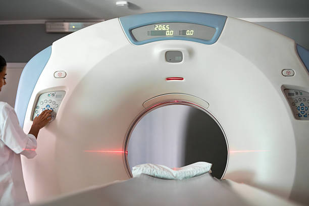
One of the most important aspects of CT investment is assessing infrastructure before signing the equipment-purchase contract.
A CT scanner contains multiple electronic systems, rotating mechanical components, and a high-power X-ray generation system.
Installation conditions can therefore directly affect performance and long-term operational reliability.
Electrical System
Facilities should confirm:
- Voltage and power-supply configuration.
- Required electrical capacity.
- Load capacity of the existing electrical system.
- Electrical distribution panel and protective equipment.
- Grounding system.
- Power-quality requirements.
- Response plan for power outages.
- Manufacturer-specific requirements.
Some systems may require power-conditioning solutions or uninterruptible power supplies for appropriate components.
However, a UPS used to protect computers or support safe system shutdown does not necessarily mean that it can maintain the CT scanner's full X-ray generation capability.
The actual scope of backup power should be clearly confirmed.
Cooling System
The X-ray tube and electronic components in a CT scanner may generate substantial heat during operation.
Depending on the model, cooling may use:
- Air-cooling solutions.
- Liquid-cooling systems.
- Chillers or heat exchangers.
- Combined configurations.
If a chiller is used, its technical requirements and compatibility with the building infrastructure should be verified.
If the equipment uses air cooling, ventilation and environmental conditions should also be evaluated.
It should not be assumed that every CT scanner uses the same type of cooling system.
Air Conditioning and Environmental Conditions
The equipment room may require control of:
- Ambient temperature.
- Humidity.
- Air circulation.
- Heat output.
- Maintenance clearance.
- Dust levels and cleanliness conditions.
Environmental conditions should remain within the limits specified by the manufacturer.
An air-conditioning system adequate for normal room use may not necessarily support the actual heat load generated by a CT scanner.
Floor Loading and Installation Space
In addition to electrical power and cooling, facilities should consider:
- Gantry weight.
- CT table loading.
- Floor load-bearing capacity.
- Equipment delivery route into the building.
- Door and corridor dimensions.
- Operating space.
- Maintenance clearance.
- Control-room location.
For hospital renovation projects, particular attention should be paid to the equipment delivery route and structural conditions of the existing building.
CT Room and Radiation Shielding
CT room design should address radiation-protection requirements for staff, patients, and surrounding areas.
Items to evaluate include:
- Gantry and table layout.
- Control area.
- Walls, doors, and shielding partitions.
- Adjacent rooms or corridors.
- Occupancy of surrounding areas.
- Expected workload.
- Ability to observe and communicate with the patient.
A single fixed lead or concrete thickness should not be applied to every CT room.
Shielding requirements should be calculated according to the building, equipment, and applicable requirements.
Radiation-safety design, assessment, and acceptance should involve appropriately qualified personnel or organizations.
7. Connecting CT with PACS and RIS
Modern CT scanners generate large volumes of image data.
Integration with data-management systems is therefore an important investment criterion.
DICOM, PACS, and RIS
DICOM (Digital Imaging and Communications in Medicine) is a standard that supports representation and exchange of medical imaging data.
PACS (Picture Archiving and Communication System) is used for image storage and communication.
RIS (Radiology Information System) supports information management and workflow within the radiology department.
A CT system may support:
- Receiving worklists through DICOM Modality Worklist.
- Transferring CT images to PACS.
- Storing reconstructed image series.
- Sending information related to performed procedures.
- Exporting radiation-dose reports where supported.
- Retrieving data for processing on other workstations.
Considerations for Exposure Data
Some CT scanners may support Radiation Dose Structured Report (RDSR) for exchanging information related to system radiation output.
This function may be useful for exposure-management and monitoring programs.
However, not all systems support the same reporting structure or scope.
During evaluation, facilities should request the supplier's DICOM Conformance Statement and verify compatibility with the existing PACS/RIS.
The presence of a network port or the ability to export DICOM images does not by itself demonstrate that the system supports all required integration workflows.
8. Quality Control and Maintenance of CT Scanners

To maintain performance and safe operation, CT scanners should be managed through an appropriate quality-control and maintenance program.
According to American College of Radiology (ACR) guidance, facilities performing CT should have a QC program supervised by a qualified medical physicist within the applicable scope.
Items to Be Checked
Evaluation items may include:
- Accuracy of CT numbers.
- Image uniformity.
- Noise level.
- Spatial resolution.
- Low-contrast detectability.
- Image artifacts.
- Accuracy of table movement.
- Alignment between the radiation field and acquired data.
- Indicators related to radiation output.
- Condition of monitors and display systems.
Testing frequency and methods should be appropriate to manufacturer instructions, the facility's quality-management program, and current requirements at the location of use.
Maintenance of the X-Ray Tube and Cooling System
The X-ray tube is one of the components that may significantly affect total ownership cost.
Facilities should clarify:
- X-ray tube warranty conditions.
- How warranty coverage is determined.
- Availability of replacement tubes.
- Replacement cost.
- System downtime during repair.
- Post-replacement testing procedures.
In addition, the cooling system, gantry, CT table, and electronic components should be maintained according to technical documentation.
Testing After Major Repairs
According to ACR QC guidance, after major repairs such as replacement of the X-ray tube or detector assembly, the system should undergo appropriate testing relevant to the repaired component.
Completion of a repair does not automatically mean that the system is ready to return to professional operation.
Relevant Technical Standard
IEC 60601-2-44 specifies particular requirements for the basic safety and essential performance of X-ray equipment for computed tomography.
As of October 2026, the consolidated version IEC 60601-2-44:2009 + Amendment 1:2012 + Amendment 2:2016 remains the published version in force according to the IEC database, while a new edition is under development.
International standards are technical references. When implementing a CT system in Vietnam, facilities should review current legal requirements relating to medical devices, radiation safety, and relevant operating conditions.
9. Ten Criteria for Selecting a CT Scanner for a Hospital
Healthcare facilities may use the following framework to compare CT systems:
| Criterion | What Should Be Evaluated? |
|---|---|
| 1. Professional requirements | Planned technical service portfolio, patient groups, and expected throughput |
| 2. Detector configuration | Number of detector rows, coverage, and acquisition capability |
| 3. X-ray tube | Power, heat-load capability, durability, and replacement options |
| 4. Image quality | Resolution, noise, reconstruction algorithms, and processing speed |
| 5. Radiation management | Optimization functions, CTDIvol/DLP indicators, and quality control |
| 6. CT table and gantry | Weight capacity, dimensions, and operational convenience |
| 7. Technical infrastructure | Electrical power, cooling, air conditioning, structural load, space, and shielding |
| 8. Data connectivity | DICOM, PACS/RIS, storage, and exposure-data management |
| 9. Maintenance service | Technical support, spare parts, X-ray tube support, and fault-response time |
| 10. Lifecycle cost | Equipment, construction, electricity, cooling, software, and maintenance |
Common CT Investment Mistakes
Comparing only slice count: Failing to evaluate physical detector configuration, acquisition speed, image quality, and software.
Overlooking cooling infrastructure: Purchasing the equipment before confirming that the room can meet environmental requirements.
Failing to budget for X-ray tube replacement: Actual ownership cost may be higher than the original estimate.
Lacking a radiation-management plan: Failing to establish a quality-control program and monitor relevant exposure indicators.
Failing to clarify software costs: Some reconstruction functions or advanced applications may require separate licenses.
Failing to assess PACS/RIS integration: Difficulties may arise when transferring, storing, or retrieving data.
Failing to account for downtime: Inadequate maintenance and technical-support arrangements may affect radiology-department operations.
10. Questions to Ask CT Suppliers Before Investment

Before selecting a system, hospitals should clarify:
1. How many physical detector rows and acquisition channels does the system have, and how is the advertised slice count determined?
2. Which professional applications are supported by the proposed configuration?
3. Which specifications and test methods are used to evaluate image quality?
4. What are the X-ray tube's heat-load characteristics, warranty conditions, and replacement costs?
5. Which exposure-optimization and exposure-monitoring functions does the system provide?
6. What specific electrical, cooling, air-conditioning, structural, and radiation-shielding requirements apply to the CT room?
7. Which reconstruction and analysis software packages are included in the quotation?
8. Which DICOM functions and PACS/RIS integration capabilities are supported?
9. Does the supplier provide site assessment, installation, acceptance testing, training, and maintenance in Vietnam?
10. How is the total multi-year investment and operating cost, including the X-ray tube, maintenance, and software, calculated?
Hospitals should ask suppliers to perform an infrastructure assessment and provide a detailed technical configuration before finalizing the investment plan.
Frequently Asked Questions About CT Scanners
What Is a CT Scanner?
A CT scanner is a system that uses X-rays and data-processing algorithms to generate cross-sectional images of internal body structures.
Is CT Different from X-Ray Imaging?
Yes. Conventional X-ray systems primarily generate two-dimensional projection images, while CT acquires data from multiple angles to reconstruct cross-sectional and volumetric images.
Is a 64-Slice CT Scanner Better Than a 16-Slice CT Scanner?
This cannot be concluded from slice count alone. Detector configuration, rotation speed, image quality, reconstruction capability, and actual intended use should also be evaluated.
Does a CT Scanner Require a Dedicated Cooling System?
It depends on the model. Some systems use air-cooling solutions, while others require liquid cooling or a chiller. Specific requirements should be confirmed according to manufacturer instructions.
Does a CT Scanner Require a Radiation-Shielded Room?
Appropriate radiation-protection measures are required according to the equipment and conditions of use. Shielding design should be assessed based on the building, workload, surrounding areas, and applicable requirements.
Are CTDIvol and DLP the Actual Radiation Dose Received by the Patient?
No. These indicators are used to describe and manage the radiation output of the CT system and are not direct measurements of the exact absorbed dose received by an individual patient.
Can a CT Scanner Connect to PACS?
Many CT systems support DICOM and PACS connectivity. However, facilities should verify connectivity functions, software licenses, and compatibility with the existing infrastructure.
Does a CT Scanner Require Periodic Maintenance?
Yes. The system should be inspected and maintained according to manufacturer instructions and the facility's quality-management program, particularly the X-ray tube, detector, gantry, cooling system, and safety functions.
Explore CT Technology and Diagnostic Imaging Solutions at Pharmedi Vietnam 2027
Investing in a CT scanner is a decision involving equipment, infrastructure, and the operational capability of the radiology department. In addition to detector configuration and image-reconstruction capability, hospitals should evaluate the X-ray generation system, radiation management, electrical power, cooling, PACS/RIS connectivity, and long-term maintenance support.
Pharmedi Vietnam 2027, taking place from September 28–30, 2027 at the Saigon Exhibition and Convention Center (SECC), Ho Chi Minh City, provides an opportunity for businesses, professionals, and healthcare organizations to meet, explore technologies, and discuss equipment solutions that support healthcare delivery.
For hospitals and diagnostic imaging centers planning to upgrade their CT systems, Pharmedi Vietnam 2027 provides an opportunity to review technology trends, explore supplier capabilities, and prepare technical requirements before selecting a solution.
Technology groups worth exploring include computed tomography systems, image-acquisition and reconstruction technologies, diagnostic data-management software, and solutions that support equipment operation.
When discussing solutions with suppliers, healthcare facilities may focus on:
- Is the detector configuration and acquisition capability appropriate for the facility's professional requirements?
- Which image-reconstruction and exposure-management technologies does the system provide?
- What are the electrical, cooling, radiation-shielding, and installation-space requirements?
- Can the system connect with the existing PACS/RIS and data infrastructure?
- Does the supplier provide site assessment, installation, training, maintenance, and X-ray tube support?
- How is the total long-term investment and operating cost determined?
Preparing these criteria helps hospitals compare solutions more effectively and develop an investment plan aligned with their actual operating scale.
For CT scanners, an effective selection should not be based only on slice count or the newest technology. It should also support image quality, radiation safety, system reliability, and sustainable long-term operation.
Dates: September 28–30, 2027
Venue: Saigon Exhibition and Convention Center (SECC), Ho Chi Minh City
Exhibition Information: https://pharmed.vn/
Product Categories: https://pharmed.vn/exhibitors/products
Visitor-registration information, participating-company lists, and activity programs will be updated according to official announcements from the Pharmedi Vietnam 2027 Organizing Committee.
This article is provided for informational purposes regarding the principles, technology, and investment criteria for CT scanners. It does not provide guidance on imaging indications, X-ray exposure settings, contrast-media use, or image interpretation for diagnosis. Installation, testing, acceptance, and operation of the system should be performed by appropriately qualified personnel in accordance with manufacturer instructions and current applicable requirements.
Related post
- Global virus vaccine race heats up, but not without controversy
- Diagnostic Imaging Equipment in Hospitals: Technology Categories and Selection Criteria
- Criteria for smart hospitals in Vietnam: What factors should medical facilities evaluate?
- Vietnam: People with Breast Cancer are getting Younger
- Vietnam: A Promising Hub for Healthcare Investment