

A magnetic resonance imaging system is a diagnostic imaging system that uses a strong magnetic field, radiofrequency waves, and signal-processing technology to generate detailed images of internal body structures. Unlike X-ray and CT systems, MRI technology does not use ionizing radiation to create images.
Magnetic Resonance Imaging (MRI) systems are now used in many hospitals and diagnostic imaging centers. Systems may differ in magnetic-field strength, magnet design, RF transmit and receive coils, gradient technology, reconstruction software, and scope of applications.
However, investing in an MRI system involves more than choosing between a 1.5 Tesla and 3 Tesla scanner. Hospitals should also evaluate image quality, RF coil configuration, data-processing capability, MRI room requirements, magnetic-field safety controls, cooling systems, and long-term maintenance capability.
In particular, although MRI does not use X-rays, the strong magnetic-field environment presents its own risks to staff, patients, and equipment brought into the scanning area.
This article focuses on the operating principles, technical components, differences among MRI configurations, and the criteria healthcare facilities should evaluate when investing in a magnetic resonance imaging system.
What Is a Magnetic Resonance Imaging (MRI) System?
An MRI system is diagnostic imaging equipment that uses the phenomenon of nuclear magnetic resonance to acquire signals from tissues and generate images through data processing.
Hydrogen atoms are widely present in water and fat within the body. The hydrogen nucleus contains a proton with magnetic properties that can interact with magnetic fields and radiofrequency energy.
The MRI system uses the main magnetic field to establish a net magnetization state among protons, then applies RF pulses to excite them and acquires signals emitted during relaxation.
The signal data are combined with spatial-encoding information generated by the gradient system to reconstruct images.
According to the U.S. National Institute of Standards and Technology (NIST), MRI generates images based on interactions among protons, magnetic fields, and radiofrequency waves, allowing information about the characteristics of internal tissues to be acquired.
Does MRI Use X-Rays?
No.
This is a fundamental difference between MRI and X-ray or CT systems.
- Digital X-ray: uses X-rays to generate projection images.
- CT: uses X-rays and data-reconstruction technology to generate cross-sectional images.
- MRI: uses magnetic fields and RF waves to acquire magnetic resonance signals.
Therefore, MRI does not expose patients to ionizing radiation through the same mechanism as X-ray or CT imaging.
However, this does not mean MRI is completely risk-free.
The static magnetic field, time-varying gradient fields, and RF energy may all create hazards that need to be controlled.
1. Operating Principle of Magnetic Resonance Imaging
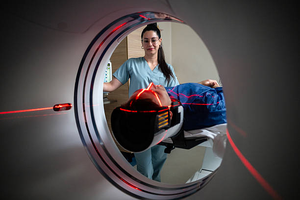
MRI image generation involves the coordinated operation of three main systems:
- Main magnetic field (B₀).
- Gradient magnetic-field system.
- RF transmit and receive system.
Main Magnetic Field B₀
The main magnetic field is generated by the MRI magnet system.
When the body is placed in the magnetic field, hydrogen protons interact with the field and establish a magnetization state that can be used for signal acquisition.
Magnetic-field strength is usually expressed in Tesla (T).
Common clinical MRI configurations include 1.5T and 3T systems. Lower-field and other specialized configurations are also available.
Magnetic-field strength is an important technical factor, but it is not the only specification that determines MRI image quality.
RF Pulses and Magnetic Resonance Signals
The system applies RF energy at an appropriate frequency to excite the proton magnetization state.
After the RF pulse ends, signals from the protons change according to the characteristics of the surrounding tissue environment and are acquired by the system.
Magnetization-relaxation characteristics, commonly described using parameters such as T1 and T2, contribute to differences in tissue contrast on MRI images.
Spatial Encoding with Gradients
If only the main magnetic field and RF waves were used, the system would not have sufficient information to determine the exact location from which a signal originated.
Gradient coils generate controlled magnetic-field variations along the spatial axes.
This allows the system to encode signal location and generate data for image reconstruction.
Image Reconstruction
MRI signals are digitized, processed, and reconstructed into images using software.
Depending on the system, functions may include:
- Multiplanar image reconstruction.
- Image acquisition using different pulse sequences.
- 3D image reconstruction.
- Noise reduction and image processing.
- Support for parallel imaging techniques.
- Advanced reconstruction algorithms.
Not all MRI systems provide the same software functions or reconstruction capabilities.
2. Components of an MRI System
A complete MRI system includes multiple electrical, magnetic, mechanical, and software components.
| Component | Primary Function |
|---|---|
| Main magnet | Generates the B₀ magnetic field |
| Gradient system | Encodes signal location spatially |
| RF coils | Transmit and/or receive magnetic resonance signals |
| RF and gradient controllers | Control pulse generation and acquisition processes |
| Patient table | Supports patient positioning and movement |
| Signal-processing system | Converts data and reconstructs images |
| Control workstation | Manages acquisition and image display |
| Cooling system | Maintains operating conditions for relevant components |
| Auxiliary equipment | Supports monitoring, communication, and safety |
| Connectivity software | Transfers images, supports storage, and integrates data |
These components may vary depending on the configuration and magnet technology.
Main Magnet
The magnet is the central component of an MRI system.
Several magnet technologies may be used:
Superconducting magnet: uses superconducting materials under appropriate cooling conditions to generate a strong and stable magnetic field.
Permanent magnet: uses magnetic materials to create a magnetic field without requiring a superconducting mechanism.
Resistive magnet: generates a magnetic field by passing electrical current through conductive coils, with its own power and cooling requirements.
Superconducting magnet technology is commonly used in widely deployed 1.5T and 3T whole-body MRI systems.
When evaluating the magnet, facilities should review:
- Magnetic-field strength.
- Field homogeneity.
- Bore size.
- Fringe-field design.
- Cooling requirements.
- Monitoring and safety mechanisms.
- Long-term maintenance capability.
Gradient System
The gradient system generates magnetic-field variations for spatial encoding.
Commonly stated specifications include:
- Gradient amplitude: gradient strength.
- Slew rate: rate of gradient change.
- Gradient linearity: linearity characteristics of the gradient.
- Cooling capability.
- Operating limits.
Gradient performance may affect the ability to implement certain imaging techniques, spatial accuracy, and other characteristics.
However, higher gradient amplitude does not automatically guarantee better image quality in every situation.
RF Coils
An RF coil is a component used to transmit or receive magnetic resonance signals depending on the configuration.
Common coil types include:
- Head coil.
- Spine coil.
- Body coil.
- Knee coil.
- Shoulder coil.
- Breast coil.
- Other specialized coils.
Multi-element receive coils may support parallel imaging techniques when compatible with the system.
When selecting a system, facilities should check the number of coils, intended applications, receive-element configuration, compatibility, and replacement cost.
The advertised number of RF channels should not be evaluated without also considering the quantity and quality of the receive coils supplied with the system.
Patient Table and Accessories
The MRI table positions the patient within the acquisition region.
Factors to evaluate include:
- Maximum load capacity.
- Travel range.
- Positioning accuracy.
- Accessibility.
- Positioning and support accessories.
- Cleaning requirements.
- Compatibility with RF coils.
Accessories brought into the MRI area must also be assessed for safety in a strong magnetic-field environment.
3. Comparing 1.5T and 3T MRI: Which Configuration Should Be Selected?
1.5 Tesla and 3 Tesla MRI are two common configurations considered when hospitals plan an MRI investment.
In principle, both use magnetic fields, RF waves, and gradients to generate images.
The difference in magnetic-field strength can affect signal characteristics, contrast, the performance of certain imaging techniques, and safety-management requirements.
Comparison of 1.5T and 3T MRI
| Criterion | 1.5T MRI | 3T MRI |
|---|---|---|
| Magnetic-field strength | 1.5 Tesla | 3 Tesla |
| Signal-to-noise ratio (SNR) | Depends on the system and acquisition method | May provide higher SNR under appropriate conditions |
| Image resolution | May support a wide range of applications | May use the SNR advantage to improve resolution |
| Acquisition speed | Depends on pulse sequence and configuration | May use the signal advantage in certain acquisition methods |
| Susceptibility artifact | Has configuration-specific characteristics | Some artifacts may be more pronounced |
| RF energy management | Requires system-specific control | May involve more complex RF-management requirements |
| Implant compatibility | Must be confirmed for each device and condition | Must be confirmed separately and should not be inferred from 1.5T compatibility |
| Investment cost | Depends on model and configuration | May be higher, but specific quotations should be compared |
| Infrastructure requirements | According to manufacturer requirements | According to manufacturer requirements and magnetic-field design |
Does 3T MRI Provide Better Images Than 1.5T MRI?
This cannot be concluded in every case.
3T MRI has the potential to provide a higher signal-to-noise ratio. Under appropriate conditions, this advantage may be used to improve resolution or adjust acquisition time.
However, 3T systems may also present challenges such as:
- Susceptibility-related artifacts.
- RF signal nonuniformity.
- RF heating-related limitations.
- Artifacts in certain imaging methods.
- Additional safety-control and device-compatibility requirements.
Therefore, 3T MRI is not automatically better than 1.5T MRI for every imaging procedure or patient group.
When selecting a system, hospitals should evaluate sample images, the planned technical service portfolio, operational capability, and the cost of each configuration.
Should a Low-Field MRI System Be Considered?
In addition to 1.5T and 3T systems, the market includes low-field MRI systems and specialized designs.
Some products emphasize flexibility, different use environments, or infrastructure requirements that differ from conventional superconducting MRI systems.
However, facilities should verify:
- Image quality.
- Coil types and software.
- Scope of applications.
- Throughput.
- Connectivity capability.
- Safety-control requirements.
- Maintenance services.
Lower magnetic-field strength does not mean that the system is suitable for every purpose or that MRI safety procedures are unnecessary.
4. What Factors Affect MRI Image Quality?

In addition to magnetic-field strength, MRI image quality is influenced by multiple system components and acquisition methods.
Signal-to-Noise Ratio (SNR)
Signal-to-Noise Ratio (SNR) reflects the relationship between useful signal and noise.
SNR is influenced by:
- Magnetic-field strength.
- RF coil design.
- Position and size of the acquisition region.
- Acquisition parameters.
- Bandwidth.
- Processing technology.
- Other technical factors.
SNR values from two promotional documents should not be compared if the measurement methods and test conditions are different.
Magnetic-Field Homogeneity
Homogeneity of the main magnetic field can affect signal quality and the ability to implement certain acquisition techniques.
MRI systems may use shimming solutions to improve magnetic-field homogeneity within the intended design range.
However, effectiveness also depends on magnet configuration, acquisition region, and other related factors.
Image Artifacts
Common MRI artifacts may include:
- Motion artifact.
- Ghosting.
- Susceptibility artifact.
- Geometric distortion.
- Interference from external RF signals.
- Artifacts related to the gradient system or RF coils.
It should not be assumed that every image-quality problem can be corrected with software.
Some issues require inspection of hardware, calibration, or room conditions.
Image-Reconstruction Technology
Some MRI systems support:
- Parallel imaging.
- Compressed sensing.
- Noise-reduction techniques.
- 3D image reconstruction.
- AI-assisted reconstruction algorithms.
- Motion-reduction or workflow-optimization tools.
However, implementation depends on hardware, RF coils, software licensing, and the scope specified by the manufacturer.
When comparing systems, facilities should clarify which functions are included in the standard configuration and which require additional purchase.
5. Important Safety Risks in the MRI Environment
MRI does not use ionizing radiation, but it has specific risks that must be managed.
According to the FDA, three major sources of risk in the MRI environment are related to:
- Strong static magnetic fields.
- Time-varying gradient fields.
- RF energy.
In addition, facilities should manage risks associated with implants, objects brought into the room, acoustic noise, and emergency situations.
Projectile Risk from Ferromagnetic Objects
A strong magnetic field can act on ferromagnetic materials.
Inappropriate objects brought into the magnetic-field area may be attracted toward the scanner, creating risks to patients, staff, and equipment.
Items requiring control may include:
- Gas cylinders.
- Carts.
- Metal tools.
- Cleaning equipment.
- Maintenance tools.
- Medical equipment not confirmed as suitable for the MRI environment.
Superconducting magnets generally maintain their magnetic field even when the scanner is not actively acquiring images.
Therefore, the absence of operating noise or a powered-off display does not mean the magnet area is safe for unrestricted equipment access.
RF Heating Risk
RF energy can cause heating during MRI.
The risk of thermal injury may increase under inappropriate conditions involving conductive materials, cables, accessories, or implanted devices.
MRI systems include RF-energy monitoring functions according to their design.
One commonly referenced indicator is the Specific Absorption Rate (SAR), which relates to the rate at which RF energy is absorbed by tissue.
However, SAR control does not eliminate every risk of burns, particularly when inappropriate local conditions are present.
Facilities should follow manufacturer safety instructions and professional procedures.
Acoustic Noise and Peripheral Nerve Stimulation
Gradient operation can generate substantial acoustic noise during image acquisition.
In addition to potential effects on hearing, time-varying magnetic fields may cause peripheral nerve stimulation under certain conditions.
Therefore, when investing in a system, facilities should evaluate:
- Protection and warning functions.
- Noise-management capability.
- Appropriate hearing-protection accessories.
- System operating limits.
- Documentation and staff training.
MRI safety should not be assessed only on whether the system includes noise-reduction technology.
6. Implant Safety and MRI Zone Management
One of the most important aspects of MRI safety management is determining which people and objects may enter the magnetic-field area.
MR Safe, MR Conditional, and MR Unsafe Classifications
According to MRI safety terminology used in FDA guidance and ASTM standards, devices or objects may be classified as:
| Classification | Meaning |
|---|---|
| MR Safe | No known hazards in MRI environments under the applicable definition |
| MR Conditional | Considered safe only when all specified conditions of use are met |
| MR Unsafe | Presents an unacceptable risk in the MRI environment |
A device labeled MR Conditional should not be assumed to be usable in every MRI scanner.
Conditions may relate to:
- Magnetic-field strength.
- Spatial magnetic-field gradient.
- RF conditions.
- SAR limits or related parameters.
- Specific use requirements.
For example, a device confirmed as suitable at 1.5T should not automatically be used at 3T.
Similarly, the absence of MRI safety information does not mean that a device is safe.
Hospitals should establish procedures to verify information and perform screening before patients or equipment enter the MRI area.
MRI Safety Zones
According to guidance from the American College of Radiology (ACR), MRI areas may be organized into four controlled zones:
- Zone I: public area without restricted access under the zoning model.
- Zone II: reception and preparation area where appropriate screening and checking activities are performed.
- Zone III: controlled-access area restricted to authorized individuals according to facility procedures.
- Zone IV: room containing the MRI magnet system.
These zones should be arranged according to the building layout, magnetic-field map, and the facility's safety program.
Facilities should particularly organize:
- Warning signage.
- Access control.
- Screening procedures.
- Staff instructions.
- Management of equipment entering the room.
- Emergency-response arrangements.
The ACR Manual on MR Safety 2026 is an updated reference for MRI facilities and includes considerations related to area control, screening, and equipment safety.
7. MRI Room Requirements: Infrastructure and Safety Design
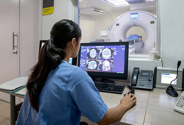
Unlike an X-ray or CT room, an MRI room should be evaluated simultaneously for magnetic-field conditions, RF interference, cooling infrastructure, structural loading, and access control.
These requirements can significantly affect implementation costs.
MRI Suite Layout
An MRI area may include:
- Magnet room.
- Control room.
- Equipment room.
- Patient reception and preparation area.
- Changing or screening area.
- Other appropriate support spaces.
The layout should support operational requirements, access safety, and emergency-response arrangements.
The project area should not be calculated only from the physical footprint of the MRI scanner.
RF Shielding and Magnetic-Field Control
MRI rooms commonly require RF shielding to reduce the effect of external electromagnetic interference on acquired signals.
This solution is commonly referred to as an RF shield or RF-shielded room.
Magnetic shielding, in contrast, is related to controlling the extent and effects of the magnetic field.
These are two different technical objectives.
| Solution | Purpose |
|---|---|
| RF shielding | Reduces radiofrequency interference affecting signal acquisition |
| Magnetic shielding | Controls the distribution and effects of the magnetic field inside and outside the equipment area |
A system with a self-shielded magnet design may still require an RF-shielded room according to manufacturer requirements.
In addition, doors, windows, electrical lines, ventilation systems, and penetrations through the shielding layer should be coordinated with the building design.
Electrical Power and Cooling Systems
Facilities should evaluate:
- Power supply and required capacity.
- Grounding system.
- Electrical protection equipment.
- Air-conditioning system.
- Temperature and humidity.
- Equipment heat load.
- Chiller or cooling system where required.
- Electrical-fault response arrangements.
It should not be assumed that every MRI system uses the same cooling configuration.
Magnet design and cooling technology determine the specific requirements.
Helium Systems and Quench Events
Some superconducting MRI systems use helium to maintain the very low temperatures required by the magnet.
A quench occurs when a superconducting magnet loses its superconducting state, causing a rapid change in stored magnetic energy and potentially releasing cold gas.
If the system uses liquid helium, uncontrolled gas release can reduce oxygen concentration in an enclosed space and create other hazards.
Therefore, depending on the design, facilities may require:
- A quench pipe to vent gas to a safe location.
- Monitoring mechanisms.
- Ventilation arrangements.
- Emergency-response procedures.
- Inspection of related components.
Some newer MRI technologies use reduced-helium designs or closed-loop cooling systems, but it should not be assumed that every model eliminates the need to assess cryogen or magnet-event risks.
In particular, stopping image acquisition, disconnecting electrical power, and intentionally initiating a magnet quench are different actions. They should not be treated as equivalent in emergency procedures.
Structural Load and Equipment Delivery
MRI magnets can be heavy and may have specialized transport requirements.
Before installation, facilities should check:
- Floor load-bearing capacity.
- Building structure.
- Door and corridor dimensions.
- Equipment delivery route.
- Lifting and rigging locations.
- Maintenance clearance.
- Safety conditions for adjacent areas.
For hospital renovation projects, an existing-site assessment should be completed before the investment plan is finalized.
8. MRI Software and PACS/RIS Connectivity
A modern MRI system includes more than the magnet and signal-acquisition hardware.
Software can significantly affect how effectively the equipment can be used.
Software Function Groups
Depending on the configuration, the system may support:
- Pulse-sequence management.
- 2D and 3D image acquisition.
- Parallel imaging techniques.
- Noise-reduction tools.
- Image reconstruction and processing.
- Patient-data management.
- Report export or data transfer.
- Advanced applications specified by the manufacturer.
When selecting a system, facilities should distinguish between:
- Software included in the standard configuration.
- Software requiring additional licensing.
- Functions that depend on RF coils or other hardware.
- Applications available only on certain models.
The same technology name should not be assumed to provide identical functionality or performance across different manufacturers.
PACS and RIS Connectivity
PACS (Picture Archiving and Communication System) supports medical image storage and communication.
RIS (Radiology Information System) supports information management and radiology workflow.
MRI systems may connect to these systems through supported DICOM functions.
Items to evaluate include:
- DICOM Storage.
- Modality Worklist.
- Procedure-status communication where supported.
- Ability to transfer data to PACS.
- Local data storage.
- Data-resend mechanisms after connection interruptions.
- Ability to export and retrieve relevant image series.
Hospitals should request the supplier's DICOM Conformance Statement and verify integration using the actual infrastructure.
The presence of a network port or DICOM export capability does not mean that the system is fully compatible with the facility's PACS/RIS.
9. Quality Control and Maintenance of MRI Systems
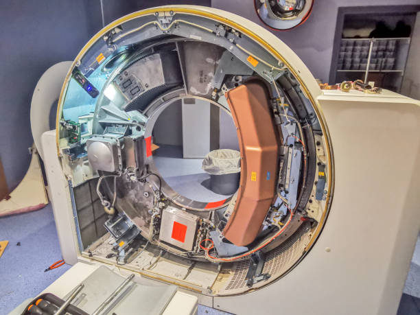
MRI systems should have an appropriate quality-control and maintenance program to maintain long-term performance.
According to ACR guidance, an MRI quality-control program should evaluate image quality, equipment performance, and relevant safety factors.
Common Quality-Control Items
| Item | Evaluation Purpose |
|---|---|
| Magnetic-field homogeneity | Evaluates B₀ field-homogeneity characteristics |
| Geometric accuracy | Evaluates dimensional and positional distortion on images |
| Slice-thickness accuracy | Checks slice-generation capability |
| Signal uniformity | Evaluates consistency of acquired signal |
| SNR | Monitors signal and noise characteristics |
| Artifact | Detects abnormal image phenomena |
| RF coils | Checks receive-coil quality and stability |
| Patient table and mechanical system | Checks positioning and movement functions |
| Safety systems | Evaluates signage, area control, and related functions |
These tests may use an MRI phantom, a specialized test object with defined characteristics used to evaluate system performance.
Testing frequency and methods should be consistent with manufacturer documentation, the quality-control program, and requirements applicable at the facility.
Magnet and Cooling-System Maintenance
For superconducting systems, maintenance programs may involve:
- Magnet status.
- Cooling system.
- Electrical and control components.
- Operational monitoring.
- Other technical components specified by the manufacturer.
It should not be assumed that every MRI system requires helium replenishment on the same schedule.
Some systems are designed to reduce the need for cryogen replenishment but still have their own technical and maintenance requirements.
RF Coil and Gradient Maintenance
In addition to the magnet, hospitals should manage:
- Condition of RF coils.
- Connectors.
- Cables.
- Amplifiers.
- Gradient system.
- Cooling system.
- Software and workstations.
After major components are repaired, appropriate testing should be performed before the system is returned to use.
Relevant MRI Technical Standards
IEC 60601-2-33:2022 specifies particular requirements for the basic safety and essential performance of magnetic resonance equipment used for medical diagnosis.
The standard addresses requirements for protecting patients and personnel working in the MRI environment.
The 2022 edition has official interpretation documents issued in 2023 and 2025.
In addition to IEC standards, facilities may refer to the ACR Manual on MR Safety 2026 when developing an MRI safety program.
These international standards and guidance documents are technical references and do not automatically constitute mandatory legal requirements in Vietnam.
10. Criteria for Selecting an MRI System for a Hospital
When evaluating MRI systems, healthcare facilities may use the following framework:
| Criterion | What Should Be Evaluated? |
|---|---|
| 1. Professional requirements | Planned technical service portfolio, patient groups, and expected throughput |
| 2. Magnetic-field strength | 1.5T, 3T, or another appropriate configuration |
| 3. Magnet design | Homogeneity, bore size, fringe field, and cooling requirements |
| 4. Gradient system | Amplitude, slew rate, performance, and operating limitations |
| 5. RF coils | Coil portfolio, signal-receive configuration, and accessories |
| 6. Image quality | SNR, resolution, artifacts, and reconstruction software |
| 7. MRI safety | Access control, RF, magnetic field, equipment, and emergency procedures |
| 8. MRI room infrastructure | Power, cooling, RF shielding, structural load, and space |
| 9. Technical service | Installation, acceptance, training, maintenance, and spare parts |
| 10. Total lifecycle cost | Equipment, construction, electricity, cooling, software, and maintenance |
Common MRI Investment Mistakes
Comparing only 1.5T and 3T: Failing to evaluate RF coils, the gradient system, image quality, and professional requirements.
Overlooking RF shielding requirements: The MRI room may not adequately control interference.
Failing to evaluate the fringe magnetic field: No appropriate plan is in place to protect adjacent areas.
Failing to assess cooling infrastructure: Additional costs or operational difficulties may arise.
Purchasing an insufficient RF coil set: The system may not support all intended applications.
Failing to establish MRI safety procedures: Screening, zoning, and equipment-control processes have not been developed.
Considering only the scanner price: Room construction, electricity, air conditioning, software, and maintenance costs are overlooked.
Failing to clarify responsibility for PACS/RIS integration: Data-management problems may arise after installation.
11. Questions to Ask MRI Suppliers Before Investment
Before purchasing a system, hospitals should clarify:
1. Is the magnetic-field strength and magnet configuration appropriate for the planned technical service portfolio?
2. What are the technical characteristics of the gradient and RF systems?
3. Which RF coils and application software packages are included in the quotation?
4. Which methods and criteria are used to evaluate image quality?
5. What specific RF shielding, fringe-field, electrical, and cooling requirements apply to the system?
6. Does the system use helium, and what cryogen-management solutions are required?
7. Does the supplier support MRI room design, site assessment, installation, and safety evaluation?
8. How does the system support PACS/RIS connectivity and data management?
9. What maintenance, fault-response, and spare-parts arrangements are available in Vietnam?
10. Which items are included in total implementation and long-term operating costs?
In particular, hospitals should ask suppliers to conduct an infrastructure assessment, provide a detailed configuration, and coordinate with the MRI room design team before the investment plan is finalized.
Frequently Asked Questions About MRI Systems
What Is a Magnetic Resonance Imaging System?
An MRI system uses magnetic fields, RF waves, and signal-processing technology to generate images of internal body structures.
Does MRI Use Ionizing Radiation?
No. Unlike X-ray and CT, MRI does not use ionizing radiation to create images. However, the technology still presents risks related to magnetic fields, RF energy, and gradients that require appropriate control.
What Is the Difference Between 1.5T and 3T MRI?
The two configurations differ in magnetic-field strength. 3T MRI may provide a higher SNR but also involves specific technical and safety requirements. Selection should be based on intended use and operational capability.
Does an MRI System Require a Shielded Room?
Different shielding and control solutions may be required, particularly RF shielding and evaluation of the extent of the magnetic field. The specific design should be based on the actual equipment and building conditions.
Does MRI Use Helium?
Many superconducting magnet systems use helium as part of the cooling technology. However, helium quantity and management requirements vary by design. Not every MRI system uses the same cooling configuration.
Can a Person with an Implant Undergo MRI?
This depends on the implant and the manufacturer's stated conditions of use. MRI safety information should be verified and appropriate professional screening performed. Safety should not be inferred solely from the device type or magnetic-field strength.
Does an MRI System Require Periodic Maintenance?
Yes. The system should be inspected and maintained according to manufacturer instructions, including the magnet, cooling system, gradients, RF system, software, and safety functions.
Explore MRI Technology and Diagnostic Imaging Solutions at Pharmedi Vietnam 2027
For hospitals and diagnostic imaging centers, investing in an MRI system involves more than choosing between 1.5T and 3T. Magnet design, RF coil quality, gradient-system performance, MRI room requirements, and maintenance capability all directly affect long-term utilization.
Pharmedi Vietnam 2027, taking place from September 28–30, 2027 at the Saigon Exhibition and Convention Center (SECC), Ho Chi Minh City, provides an opportunity for businesses, professionals, and healthcare organizations to meet, explore technologies, and discuss solutions that support healthcare delivery.
For facilities planning to invest in or upgrade MRI systems, Pharmedi Vietnam 2027 provides an opportunity to review diagnostic imaging technology trends, explore suppliers, and define the technical requirements that should be evaluated before selecting equipment.
Relevant solution groups include magnetic resonance systems, image-acquisition and processing technologies, diagnostic data-management software, and solutions that support equipment operation.
When exploring MRI systems, hospitals may focus on:
- Which 1.5T or 3T MRI configuration is appropriate for the planned technical service portfolio and expected throughput?
- What are the technical characteristics of the magnet, gradient system, and RF coils?
- Which image-reconstruction and processing software applications are integrated?
- What RF shielding, magnetic-field, electrical, and cooling requirements apply to the MRI room?
- Does the system support PACS/RIS connectivity and image-data management?
- Does the supplier provide site assessment, installation, training, and maintenance support in Vietnam?
- How are total investment, MRI room construction, and long-term operating costs determined?
Preparing these criteria helps healthcare facilities evaluate solutions proactively and develop an investment plan aligned with infrastructure conditions, professional requirements, and actual operational capability.
For MRI systems, an effective selection should support image quality, magnetic-field safety, system integration, and stable operation throughout the equipment lifecycle.
Dates: September 28–30, 2027
Venue: Saigon Exhibition and Convention Center (SECC), Ho Chi Minh City
Exhibition Information: https://pharmed.vn/
Product Categories: https://pharmed.vn/exhibitors/products
Visitor-registration information, participating-company lists, and activity programs will be updated according to official announcements from the Pharmedi Vietnam 2027 Organizing Committee.
This article is provided for informational purposes regarding the operating principles, system components, safety requirements, and selection criteria for magnetic resonance imaging systems. It does not provide guidance on imaging indications, pulse-sequence settings, implant assessment for individual patients, or image interpretation for diagnosis. Installation, acceptance, safety testing, and operation of the system should be performed by appropriately qualified personnel in accordance with manufacturer instructions and current applicable requirements.
Related post
- Introducing Vietnam - Korea Joint Venture Medical Company (VIKOMED) - The Gold Sponsor for Pharmedi Vietnam 2024
- TRIỂN LÃM TRỰC TUYẾN Y TẾ QUỐC TẾ VIỆT NAM – PHARMED & HEALTHCARE VIETNAM 2021
- Nơi trình diễn của các sản phẩm công nghệ Triển lãm Y tế Quốc tế Việt Nam lần thứ 13 – Pharmed & Healthcare Vietnam
- [Video] PHARMEDI 2019: Taiwanese businesses
- Doanh nghiệp Việt trước cơ hội xuất khẩu khẩu trang đi châu Âu, châu Mỹ