


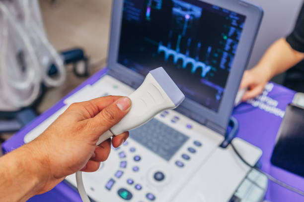
A color Doppler ultrasound system is a diagnostic imaging device that uses ultrasound waves to visualize internal body structures while applying the Doppler effect to display information about blood-flow motion. This technology combines two-dimensional ultrasound imaging with color-coded Doppler data, supporting qualified healthcare personnel in assessing flow characteristics within blood vessels and related structures.
Color Doppler ultrasound systems are currently used across many specialties, including cardiology, diagnostic imaging, obstetrics and gynecology, and vascular assessment. Depending on the configuration, the equipment may integrate multiple imaging modes, specialized transducers, measurement software, and data-management functions.
However, selecting a color ultrasound system involves more than comparing display size or the number of available imaging modes. Hospitals and clinics should also evaluate B-mode image quality, Doppler sensitivity, signal-acquisition capability, compatible transducers, processing software, safety controls, and maintenance services.
This article focuses on the operating principles, Doppler technologies, system components, and technical criteria that should be considered when investing in a color Doppler ultrasound system for a healthcare facility.
What Is a Color Doppler Ultrasound System?
A color Doppler ultrasound system is an ultrasound system capable of using the Doppler effect to acquire information related to motion and display flow data in color over the ultrasound image.
In conventional ultrasound, the transducer emits high-frequency sound waves into the body and receives returning echoes from tissues. The system processes these signals to generate images of anatomical structures.
With Doppler ultrasound, the equipment also analyzes frequency changes in the returning waves from moving structures, particularly blood cells.
From this data, the software can generate color-coded images or Doppler spectral waveforms depending on the selected mode.
According to RadiologyInfo, Doppler ultrasound may support assessment of blood-flow movement in arteries and veins and provide information about flow direction and velocity within the technical limits of the method.
Does Color Doppler Ultrasound Use X-Rays?
No.
Doppler ultrasound uses sound waves and does not use ionizing radiation like X-ray or CT imaging.
However, ultrasound still introduces acoustic energy into the body. Operators should therefore follow appropriate safety principles, particularly when using Doppler modes with different acoustic-output characteristics.
1. Operating Principle of Color Doppler Ultrasound
The Doppler principle is based on changes in wave frequency when there is relative motion among the transmitter, reflecting object, and receiver.
In medical ultrasound, the transducer sends sound waves into the area being examined. When the waves interact with moving blood cells, the frequency of the returning signal may change.
This change is known as the Doppler frequency shift.
The system analyzes the signal to generate data representing flow characteristics.
How Color Doppler Images Are Generated
The technical principle can be summarized in four stages:
1. Ultrasound transmission: The transducer emits ultrasound pulses into the area being examined.
2. Echo acquisition: Returning signals from tissues and moving structures are received by the transducer.
3. Doppler signal processing: The system analyzes information related to motion, direction, and signal characteristics according to the selected mode.
4. Data display: Doppler information is color-coded and overlaid on the background ultrasound image.
This allows the operator to observe anatomical structures together with flow information on the display.
What Do Red and Blue Mean on a Doppler Image?
This is a commonly misunderstood point.
In a typical display convention, two color groups are used to distinguish the direction of blood-flow motion relative to the transducer. They do not directly identify arterial blood versus venous blood.
For example, with a commonly used color scale:
- Red may represent flow moving toward the transducer.
- Blue may represent flow moving away from the transducer.
However, the color scale may be reversed or modified according to display settings.
Therefore, red should not automatically be interpreted as arterial flow or blue as venous flow.
Colors should be interpreted according to the active color scale, transducer position, and other professional information.
Factors Affecting Doppler Signals
The ability to detect flow depends on multiple factors:
- Ultrasound frequency.
- Angle between the ultrasound beam and direction of flow.
- Flow-motion characteristics.
- Depth of the region being examined.
- Transducer sensitivity.
- Signal-processing capability.
- Appropriate technical settings.
Angle dependence is an important limitation of Doppler ultrasound, particularly for velocity measurements. Doppler measurements should not be assumed to be independent of the insonation angle.
2. Imaging Modes on a Color Doppler Ultrasound System
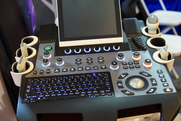
A color Doppler ultrasound system commonly combines multiple imaging modes.
These modes serve different purposes and should not be considered equivalent.
B-Mode – Two-Dimensional Ultrasound
B-mode (Brightness Mode) generates grayscale ultrasound images.
The system uses returning echoes to display anatomical structures at different brightness levels.
This is an important baseline mode for visualizing tissues and organs and locating areas for Doppler assessment.
Color Doppler
Color Doppler encodes flow information using color and overlays it on the B-mode image.
This mode may support visualization of:
- Presence of a flow signal.
- Direction of motion relative to the transducer.
- Distribution of flow within the region being examined.
- Changes in the signal over time.
Display characteristics depend on the color scale, signal quality, processing system, and imaging conditions.
Power Doppler
Power Doppler displays information based on the energy of the Doppler signal.
This mode often provides greater sensitivity for detecting certain weak flow signals compared with conventional Color Doppler.
However, standard Power Doppler does not provide flow-direction information in the same way as Color Doppler.
Some systems may include directional Power Doppler variants, depending on the manufacturer's technology.
PW Doppler – Pulsed Wave Doppler
Pulsed Wave Doppler (PW Doppler) uses ultrasound pulses to acquire motion information from a defined sample volume.
The data are commonly displayed as a velocity spectrum over time.
This mode may support measurement of flow-related parameters within an appropriate scope of use.
One technical limitation of PW Doppler is aliasing, which may occur when the Doppler frequency shift exceeds the sampling range the system can accurately represent under the corresponding conditions.
CW Doppler – Continuous Wave Doppler
Continuous Wave Doppler (CW Doppler) uses continuous transmission and reception to acquire motion signals.
This mode can support assessment of higher velocities without the same aliasing limitation mechanism as PW Doppler.
However, CW Doppler does not isolate a specific sample volume at a defined depth in the same way as conventional PW Doppler.
Not every color Doppler ultrasound system includes CW Doppler in its standard configuration.
Comparison of Ultrasound Modes
| Mode | Primary Function | Key Consideration |
|---|---|---|
| B-mode | Generates grayscale structural images | Not a dedicated mode for assessing flow |
| Color Doppler | Displays flow information in color | Depends on flow direction, angle, and color scale |
| Power Doppler | Displays Doppler signal energy | Sensitive to certain low-flow signals; standard mode does not show direction |
| PW Doppler | Records spectral data from a sample volume | May be affected by aliasing |
| CW Doppler | Records continuous Doppler spectral data | Suitable for certain high velocities but does not isolate a specific depth |
| M-mode | Displays structural motion over time | Supports motion assessment and is not equivalent to Color Doppler |
During procurement, facilities should determine which modes are included as standard and which require additional software or transducers.
3. How Does Color Doppler Ultrasound Differ from 2D, 3D, and 4D Ultrasound?
In the market, terms such as 2D ultrasound, color ultrasound, 3D ultrasound, and 4D ultrasound are often used within the same product category.
However, they do not describe the same technical characteristic.
2D ultrasound: generates two-dimensional images of anatomical structures.
Color Doppler ultrasound: uses color to represent flow information on the ultrasound image.
3D ultrasound: reconstructs ultrasound data into volumetric or three-dimensional displays.
4D ultrasound: generally refers to real-time or near-real-time display of volumetric data, depending on the system.
An ultrasound system may support 2D, Color Doppler, and 3D/4D functions at the same time if equipped with appropriate hardware and software.
Conversely, the ability to display 3D images does not automatically mean the system includes a complete range of Doppler modes.
Is Every Ultrasound Machine with a Color Display a Color Doppler System?
No.
A color display and Color Doppler functionality are two different features.
A device with a color LCD screen may still support only basic ultrasound modes.
When reviewing the technical configuration, facilities should verify Color Doppler, Power Doppler, PW Doppler, and CW Doppler functions directly rather than relying only on the commercial term “color ultrasound machine.”
4. Components of a Color Doppler Ultrasound System
A color Doppler ultrasound system commonly includes multiple electronic, software, and accessory components.
| Component | Primary Function |
|---|---|
| Ultrasound transducer | Transmits and receives ultrasound signals |
| Pulse-generation and control system | Controls ultrasound-wave transmission |
| Signal receiver and processor | Amplifies, digitizes, and processes returning data |
| Beamformer | Coordinates signals from transducer elements |
| Doppler processor | Analyzes motion-related information |
| Image processor | Generates B-mode images and display overlays |
| Display and control panel | Displays images and supports system operation |
| Memory and software | Stores results, manages data, and supports measurement functions |
| Connectivity ports | Connect transducers, network systems, and peripheral devices |
| Power supply and battery, where applicable | Provide power to the system |
Ultrasound Transducer
The transducer is the component that directly transmits and receives sound waves.
Many transducers use piezoelectric elements to convert electrical energy into ultrasound waves and vice versa.
Transducer performance depends on its design, frequency, element structure, and how the system processes the signal.
Signal Processor
The electronic system receives signals from the transducer and performs functions such as amplification, digitization, filtering, and signal coordination.
In color Doppler ultrasound, processing also involves analyzing motion-related changes to generate Doppler data.
Specifications such as the number of processing channels or frame rate may provide information about system design, but they are not sufficient to independently determine image quality.
According to recommendations from the American Institute of Ultrasound in Medicine (AIUM), ultrasound-system selection should be based on the overall performance of the system together with its transducers and its ability to meet actual professional requirements.
5. Common Types of Color Doppler Ultrasound Transducers
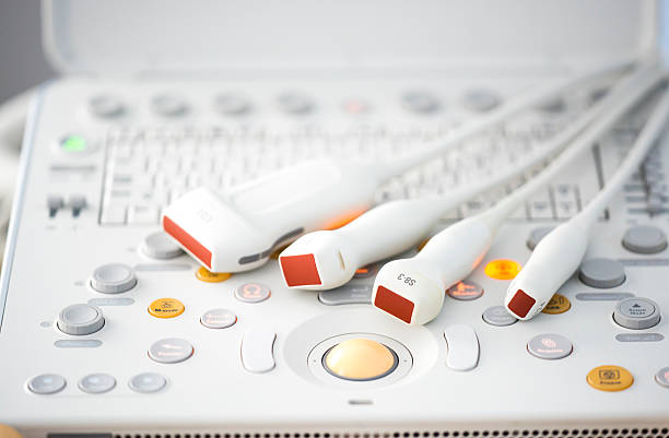
Transducer selection directly affects how effectively an ultrasound system can be used.
A system may offer many functions yet still fail to meet a facility's professional requirements if the appropriate transducers are not included.
Convex Transducer
A convex transducer has a curved transmitting and receiving surface and is commonly used for applications requiring assessment of relatively deep structures.
Common characteristics include:
- Curved fan-shaped field of view.
- Imaging coverage suitable for many general applications.
- Frequency range according to transducer design.
Linear Transducer
A linear transducer commonly provides a rectangular field of view and is used in many applications involving superficial structures.
Application groups may include:
- Peripheral vessels.
- Thyroid.
- Soft tissues.
- Musculoskeletal structures.
- Other superficial structures.
Phased Array Transducer
A phased array transducer commonly has a small contact footprint and produces a fan-shaped field of view.
This type of transducer is widely used in appropriate cardiac ultrasound systems.
Depending on the configuration, the system may support dedicated Doppler modes for cardiovascular assessment.
Endocavitary Transducer
Some systems also support endocavitary transducers for appropriate specialized applications.
These transducers have specific requirements related to:
- Shape and size.
- Frequency range.
- Intended-use scope.
- Protective covers.
- Cleaning and disinfection.
- Compatibility with reprocessing chemicals.
Transducer Selection by Application
| Transducer Type | Technical Characteristics | Common Application Groups |
|---|---|---|
| Convex | Curved surface with a wide field of view at depth | General ultrasound, abdominal imaging, and other appropriate applications |
| Linear | Flat surface, commonly supports superficial imaging | Peripheral vessels, thyroid, and soft tissues |
| Phased Array | Small footprint with a fan-shaped field of view | Cardiology and specialized applications |
| Endocavitary | Designed for intracavitary examinations | Gynecology and other specified applications |
One important principle is that higher-frequency transducers generally provide better resolution for superficial structures but may have reduced penetration, while lower-frequency transducers are often more suitable for deeper imaging.
However, image quality also depends on transducer design and the system's processing capability.
Transducers should not be selected solely on the basis of the highest frequency stated in the specification sheet.
6. Factors That Determine Doppler Image Quality
Color Doppler image quality depends on multiple interacting factors and not only on B-mode image resolution.
Resolution and Penetration
Two characteristics to evaluate are:
- Spatial resolution: ability to distinguish structures located close to one another.
- Penetration: ability to acquire useful signals at appropriate depths.
Transducer characteristics, signal-processing capability, and tissue properties may all affect results.
Color Doppler Sensitivity
Doppler sensitivity relates to the system's ability to detect and display motion signals under defined conditions.
When evaluating the system, facilities should consider:
- Ability to display weak flow signals.
- Level of color noise.
- Stability of the color region.
- Alignment with the B-mode image.
- Display performance under different examination conditions.
A system that displays more color signals does not automatically provide better Doppler quality.
Displayed data should be evaluated together with the system's ability to distinguish genuine signals from noise.
Frame Rate
Frame rate represents the number of frames displayed per unit of time.
This specification may affect how smoothly moving structures and flow are visualized.
However, frame rate is commonly related to imaging depth, Doppler region size, and acquisition method.
Therefore, maximum frame rates from two systems should not be compared if the measurement conditions are different.
Image-Processing Technologies
Some systems integrate technologies such as:
- Tissue Harmonic Imaging: uses harmonic signals to generate images under appropriate conditions.
- Spatial Compound Imaging: combines data from multiple directions to improve certain image characteristics.
- Speckle Reduction: processing intended to reduce speckle noise in images.
- Doppler Processing: algorithms that support processing of flow signals.
- AI-assisted Imaging: functions that may support recognition, measurement, or workflow optimization within the manufacturer's stated scope.
These features should be distinguished from actual system performance.
The presence of AI technology or multiple processing algorithms does not by itself demonstrate that a device will produce more accurate images in every application.
During a demonstration, facilities should request image-quality evaluation using the exact transducer types and examination groups intended for actual use.
7. Common Color Doppler Ultrasound System Configurations
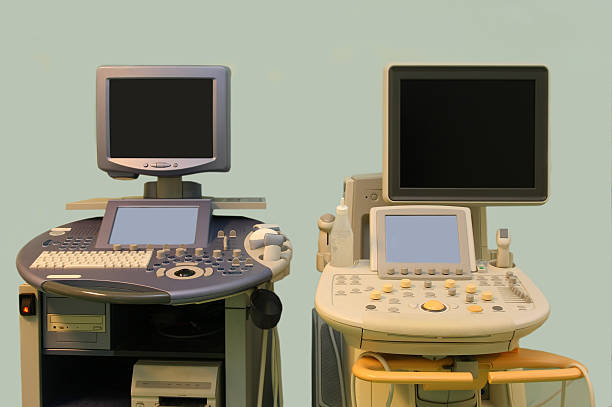
Doppler ultrasound systems are available in several configurations.
Cart-Based Ultrasound System
Cart-based ultrasound systems are commonly used in dedicated ultrasound examination rooms.
Potential operational advantages may include:
- Large display.
- Dedicated control panel.
- Multiple transducer ports.
- Storage space for accessories.
- Ability to integrate software functions.
However, size and weight may affect mobility between clinical areas.
Portable Ultrasound System
A portable color Doppler ultrasound system is designed for easier transport or use in multiple locations.
When selecting one, facilities should evaluate:
- Weight.
- Battery runtime.
- Transducer connectivity.
- Display quality.
- Range of Doppler modes.
- Operational stability.
- Cleanability.
- Cart or stand accessories.
It should not be assumed that every portable system provides the same complete functionality as a dedicated cart-based system.
Handheld Ultrasound System
Some modern ultrasound systems use transducers connected to compatible display devices.
These devices may be suitable for certain point-of-care ultrasound (POCUS) activities within the manufacturer's stated intended use.
However, facilities should verify:
- Does the system support Color Doppler?
- Does it support PW or CW Doppler?
- Is image quality appropriate for the intended use?
- Does it require network connectivity or a software subscription?
- How are data stored and protected?
Portability does not mean that the system includes a complete range of advanced Doppler functions.
8. Ultrasound Safety and Infection Control
Although ultrasound does not use ionizing radiation, the system should still comply with appropriate safety and quality-control requirements.
The ALARA Principle in Ultrasound
ALARA (As Low As Reasonably Achievable) is the principle of maintaining energy exposure at a reasonably low level while still obtaining the required professional information.
Commonly displayed ultrasound indices include:
- Thermal Index (TI): an index related to the potential for temperature increase.
- Mechanical Index (MI): an index related to the potential mechanical effects of ultrasound.
These are indicators that support safety management. They are not direct measurements of actual temperature in every tissue or absolute predictors of biological effects.
According to the FDA and AIUM, operators should consider acoustic-output levels and exposure duration to support prudent ultrasound use, particularly in patient groups requiring additional attention.
Cleaning and Disinfection of Ultrasound Transducers
Ultrasound transducers come into direct contact with patients and therefore require an appropriate reprocessing procedure.
According to AIUM guidance updated in 2025:
- External transducers that contact intact skin generally require cleaning and low-level disinfection according to an appropriate procedure.
- Endocavitary transducers that contact mucous membranes require cleaning and high-level disinfection according to guidance, together with an appropriate protective cover.
- Transducers used during procedures should be managed according to the level of tissue contact, aseptic requirements, and manufacturer instructions.
A single chemical or disinfection method should not be used for every transducer.
Incompatible chemicals may damage the transducer surface, acoustic lens, cable, or connector.
Relevant Technical Safety Standard
IEC 60601-2-37:2024 specifies particular requirements for the basic safety and essential performance of ultrasonic medical diagnostic and monitoring equipment.
This standard does not generally apply to therapeutic ultrasound equipment.
When evaluating a color Doppler ultrasound system, facilities may request technical documentation and information from the supplier regarding conformity with relevant standards.
Reference to an international standard does not mean that all provisions of that standard automatically constitute mandatory legal requirements in Vietnam.
9. Data Storage and Connectivity of Color Doppler Ultrasound Systems
For facilities pursuing digital workflows, ultrasound image and data management is an important investment criterion.
Depending on the model, the equipment may support:
- Still-image storage.
- Video or cine-loop storage.
- Report export.
- Network data transfer.
- Connectivity with centralized storage systems.
- Patient-information management.
- Dedicated measurement and reporting functions.
DICOM and PACS
DICOM (Digital Imaging and Communications in Medicine) is a standard used to represent and exchange medical imaging data.
PACS (Picture Archiving and Communication System) supports image storage, transfer, and retrieval.
An ultrasound system may support DICOM functions such as:
- DICOM Storage: transfers images to a compatible system.
- Modality Worklist: receives worklists and related information.
- DICOM Print: supports printing through compatible systems.
- DICOM Structured Report: exchanges structured reporting information within the supported scope.
Not every ultrasound system supports all of these functions.
Some functions may also require additional software licenses.
HIS/RIS Integration and Result Management
If a hospital needs to connect the ultrasound system with its information-management infrastructure, it should clarify:
- Does the system support DICOM?
- Which DICOM functions are licensed?
- In which formats can images and videos be exported?
- How is patient information synchronized?
- Does the system support Worklist?
- How long are data stored locally?
- Can images be resent after a network interruption?
- Does the supplier support PACS/HIS/RIS integration?
The presence of USB, LAN, or Wi-Fi connectivity does not mean that the ultrasound system will automatically provide full connectivity with PACS or hospital information systems.
Facilities should request the DICOM Conformance Statement and perform connectivity testing using the actual infrastructure.
10. Quality Control and Maintenance of Color Doppler Ultrasound Systems
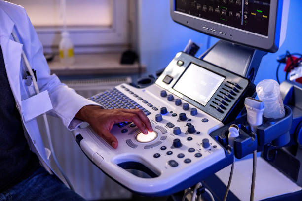
Ultrasound systems should be inspected and maintained to preserve imaging performance, signal quality, and safety during use.
Transducer Inspection
The transducer is a component that requires particular attention.
Inspection items may include:
- Condition of the transducer surface.
- Acoustic lens.
- Cable.
- Connector.
- Cracks or damage.
- Signal stability.
- Loss of transmitting or receiving elements.
- Image and Doppler quality.
A damaged transducer may not only present mechanical issues but may also reduce image quality or measurement performance.
Image-Performance Testing
Depending on the quality-control program, facilities may use an ultrasound phantom to assess characteristics such as:
- Visualization depth.
- Resolution.
- Uniformity.
- Contrast discrimination.
- Abnormal image artifacts.
- Other appropriate indicators.
Testing should be performed using appropriate methods and equipment.
Main-System Maintenance
In addition to the transducer, components that may require management include:
- Display monitor.
- Keyboard and control panel.
- Connectivity ports.
- Power system.
- Battery for mobile equipment.
- Cooling fans.
- Printer or peripheral accessories.
- Software and data storage.
According to AIUM guidance on ultrasound-facility quality management, equipment should be maintained in appropriate operating condition and periodic quality-control records should be kept.
The specific maintenance schedule should be based on manufacturer instructions, level of use, and the facility's equipment-management procedures.
11. Ten Criteria for Selecting a Color Doppler Ultrasound System for a Healthcare Facility
Hospitals and clinics may use the following framework to compare systems:
| Criterion | What Should Be Evaluated? |
|---|---|
| 1. Professional requirements | Cardiology, vascular, obstetrics and gynecology, general imaging, or other applications |
| 2. Doppler modes | Color, Power, PW, CW, and related functions |
| 3. Image quality | Resolution, penetration, Doppler sensitivity, and noise |
| 4. Transducers | Types, frequency ranges, durability, and compatibility |
| 5. Processing technology | Beamforming, image processing, measurement functions, and software |
| 6. User interface | Display, control panel, workflow, and operational management |
| 7. Storage and connectivity | DICOM, PACS, HIS/RIS, images, videos, and reports |
| 8. Safety and infection control | TI/MI, transducers, consumables, cleaning, and disinfection |
| 9. Technical service | Installation, training, warranty, spare parts, and maintenance |
| 10. Lifecycle cost | System price, transducers, software, consumables, and repairs |
Common Selection Mistakes
Looking only at the number of Doppler modes: Failing to evaluate actual sensitivity and signal quality.
Selecting a system based on processing-channel count: Overlooking the overall performance of the system together with its transducers.
Failing to confirm CW Doppler: Purchasing a system for advanced applications and later finding that the required function is not included in the configuration.
Purchasing too few transducers: The system may not support the planned technical service portfolio.
Focusing only on a large display: Failing to evaluate resolution, penetration, and Doppler visualization capability.
Failing to verify DICOM: The system may be difficult to integrate with the hospital's image-storage infrastructure.
Overlooking transducer-replacement costs: Total operating costs may exceed the original plan.
Failing to evaluate disinfection workflows: The supplied transducers may not be compatible with the facility's existing reprocessing methods.
12. Questions to Ask Color Doppler Ultrasound Suppliers
Before making an investment decision, healthcare facilities should clarify:
1. Which professional applications and Doppler modes does the system support?
2. Are Color Doppler, Power Doppler, PW, and CW included in the supplied configuration?
3. Which transducer types are included, and can additional transducers be added later?
4. Under which methods and test conditions is image quality evaluated?
5. Does the system support dedicated image-processing technologies and measurement functions?
6. Can the system store and export images and videos or connect with PACS/RIS?
7. Which software functions require additional licenses?
8. What cleaning, disinfection, and storage requirements apply to the transducers?
9. Does the supplier provide equipment demonstrations, training, maintenance, and transducer replacement support in Vietnam?
10. How is the total multi-year cost of ownership, including transducers, software, and accessories, determined?
Facilities should request a demonstration using the exact system configuration, transducers, and software they intend to purchase, with direct evaluation by the healthcare professionals who will use the equipment.
This approach helps assess system suitability more clearly than comparing specification sheets alone.
Frequently Asked Questions About Color Doppler Ultrasound Systems
What Is a Color Doppler Ultrasound System?
It is an ultrasound system that uses the Doppler effect to acquire information about motion, particularly blood flow, and display that information in color together with the ultrasound image.
Is Every Color Ultrasound Machine a Color Doppler System?
Not necessarily. A system with a color display does not automatically support Color Doppler. The stated technical functions should be confirmed directly in the product documentation.
How Does Color Doppler Ultrasound Differ from 2D Ultrasound?
2D ultrasound primarily generates grayscale structural images, while Color Doppler adds color-coded flow information.
Can Color Doppler Distinguish Arteries from Veins Based on Red and Blue Colors?
No. Color alone cannot be used for this purpose. The colors commonly represent flow direction relative to the transducer according to the active color scale and do not directly identify an artery or vein.
Are Color Doppler and Power Doppler the Same?
No. Color Doppler provides information related to flow direction, while standard Power Doppler primarily represents Doppler signal energy and is often more sensitive to certain low-flow signals.
Does a Color Doppler Ultrasound System Need Multiple Transducers?
This depends on professional requirements. Convex, Linear, Phased Array, and endocavitary transducers support different application groups. Facilities should select transducers according to the planned technical service portfolio.
Can a Color Doppler Ultrasound System Connect to PACS?
Many systems support PACS connectivity through DICOM. However, the functions, software licenses, and model-specific compatibility should be confirmed.
Does a Color Doppler Ultrasound System Require Periodic Maintenance?
Yes. Facilities should inspect transducers, electronic systems, image quality, software, and related components according to manufacturer instructions and the facility's equipment-management program.
Explore Color Doppler Ultrasound Technology and Diagnostic Solutions at Pharmedi Vietnam 2027
For hospitals and clinics, investment in a color Doppler ultrasound system should be evaluated across multiple factors, from image quality, Doppler sensitivity, and specialized transducers to analysis software, data connectivity, and technical service.
Pharmedi Vietnam 2027, taking place from September 28–30, 2027 at the Saigon Exhibition and Convention Center (SECC), Ho Chi Minh City, provides an opportunity for businesses, professionals, and healthcare organizations to meet, explore technologies, and discuss equipment solutions that support healthcare delivery.
For facilities planning to invest in or upgrade ultrasound systems, Pharmedi Vietnam 2027 provides an opportunity to review diagnostic imaging technology trends, explore supplier capabilities, and prepare technical requirements before selecting equipment.
Relevant solution groups include diagnostic ultrasound systems, Doppler technologies, specialized transducers, image-processing software, and ultrasound data-management solutions.
When discussing solutions with suppliers, hospitals and clinics may focus on:
- Which Color Doppler, Power Doppler, PW, and CW modes does the system support?
- Are image quality and Doppler sensitivity appropriate for the facility's professional requirements?
- Which transducer types are supplied, and what options are available for future expansion?
- Does the system support measurements, image and video storage, and PACS/RIS connectivity?
- What cleaning, disinfection, and maintenance requirements apply to the transducers and related components?
- Does the supplier provide equipment demonstrations, training, and spare-parts support in Vietnam?
- How is total long-term investment and operating cost determined?
Preparing these criteria helps healthcare facilities take a more proactive approach when reviewing technologies, comparing systems, and developing an investment plan aligned with their actual operating scale.
For color Doppler ultrasound systems, an effective selection should be based on the ability to meet diagnostic requirements, signal quality, integration between the system and transducers, and reliable long-term operation rather than only the number of features or initial purchase price.
Dates: September 28–30, 2027
Venue: Saigon Exhibition and Convention Center (SECC), Ho Chi Minh City
Exhibition Information: https://pharmed.vn/
Product Categories: https://pharmed.vn/exhibitors/products
Visitor-registration information, participating-company lists, and activity programs will be updated according to official announcements from the Pharmedi Vietnam 2027 Organizing Committee.
This article is provided for informational purposes regarding the operating principles, technologies, and selection criteria for color Doppler ultrasound systems. It does not provide instructions for performing measurements, setting ultrasound parameters for individual patients, or interpreting images for diagnosis. Equipment use and evaluation of results should be performed by appropriately qualified personnel according to manufacturer instructions and facility procedures.
Related post
- According to MA. Dinh Anh Nguyen, despite the fact that Vietnam’s GDP is recently assessed at a normal level, the investment in medical & pharmaceuticals fields shows a stark contrast…
- Medical Device Import Licenses: When Are They Required and What Should Businesses Prepare?
- [VIDEO] PHARMED & HEALTHCARE VIETNAM 2019 - Highlight
- Triển lãm Y tế Quốc tế lần thứ 14 tại Việt Nam- Diễn đàn giao thương các doanh nghiệp Đức